





















Description
Shining 3D · Wireless scanner + photogrammetry
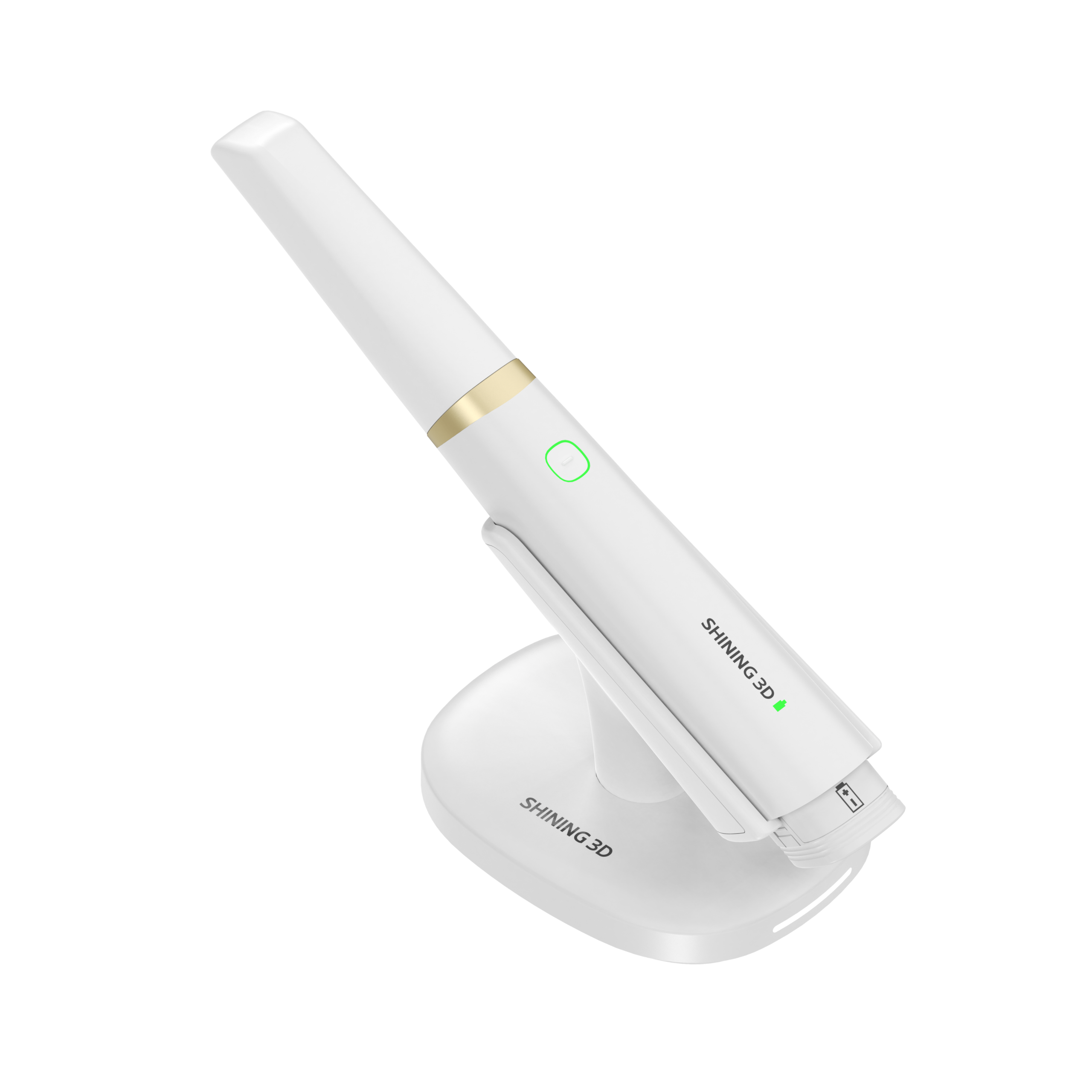
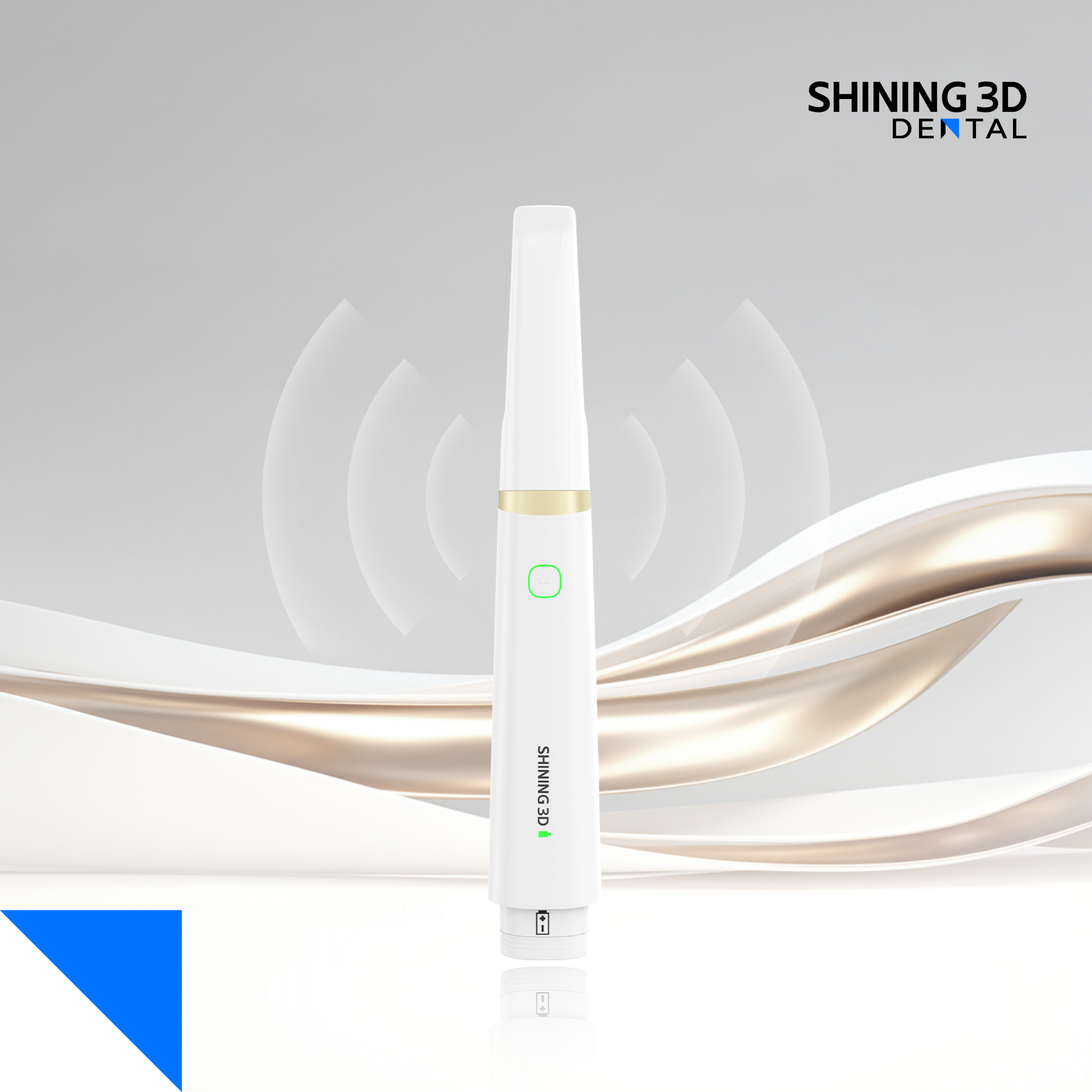
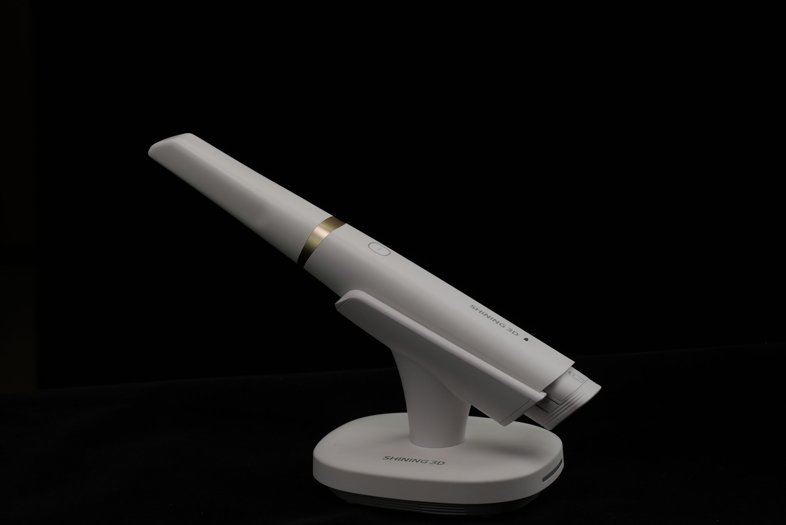
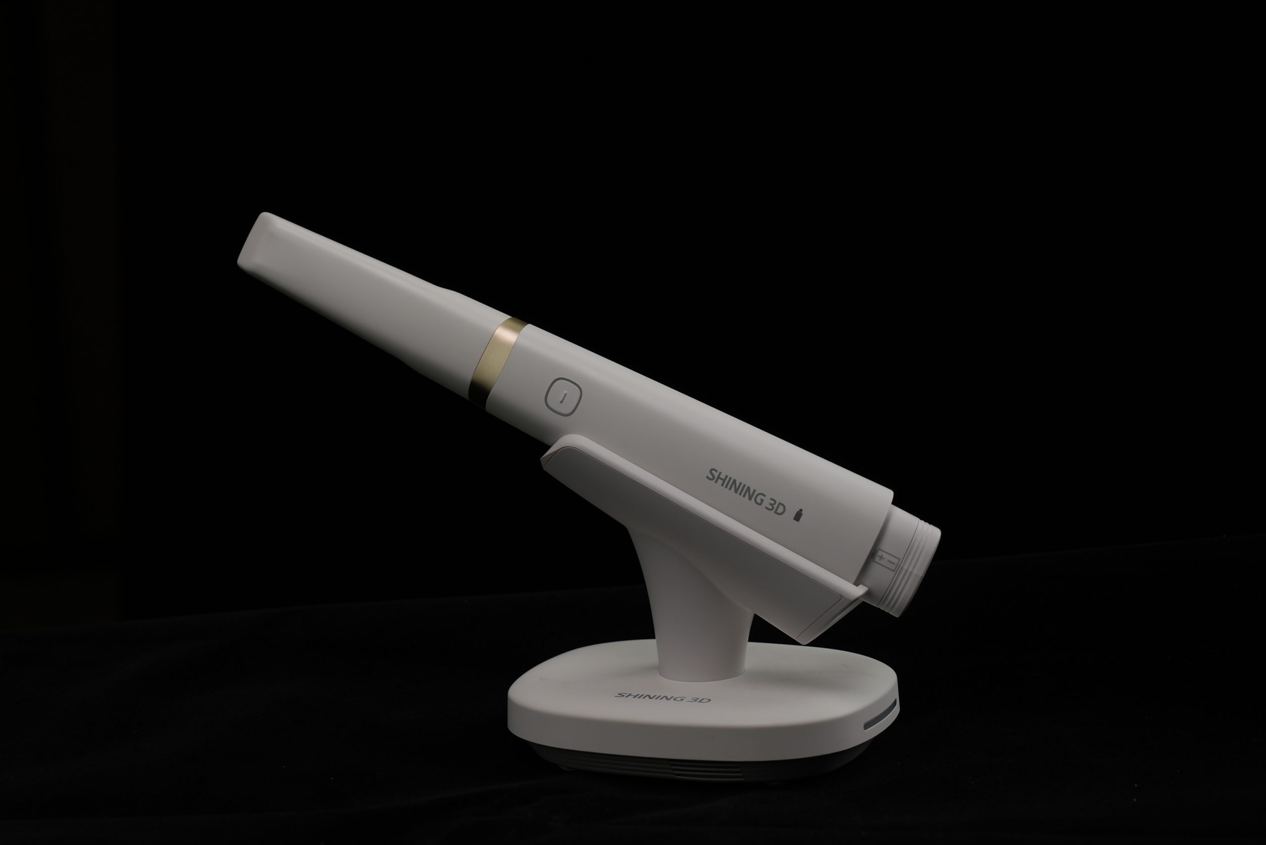
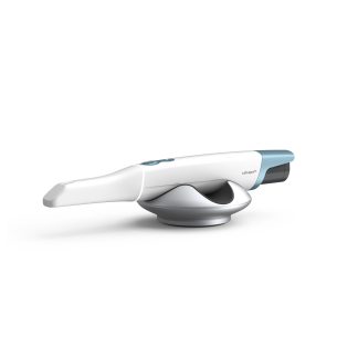
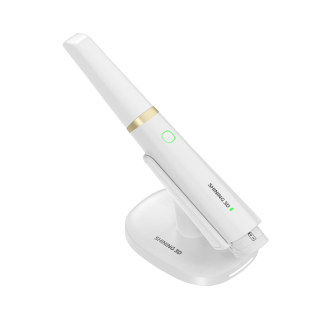
Shining 3D Aoralscan Elite Wireless
Full-arch implant photogrammetry, now without the cable.
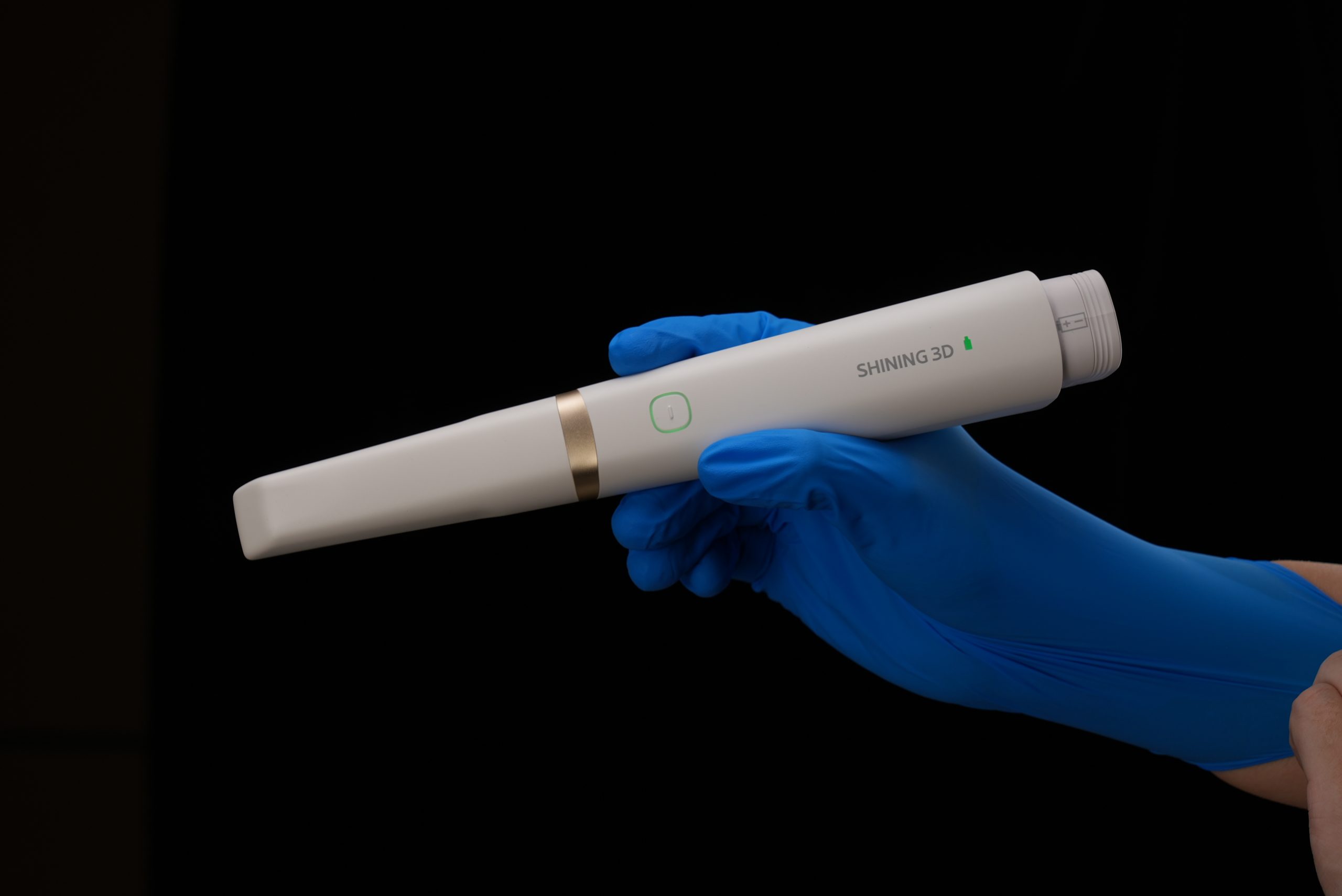
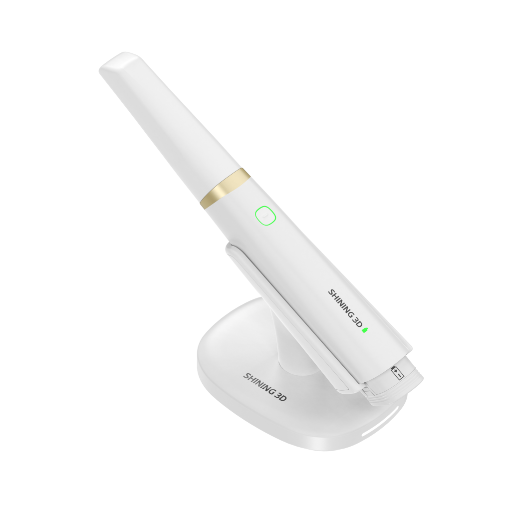
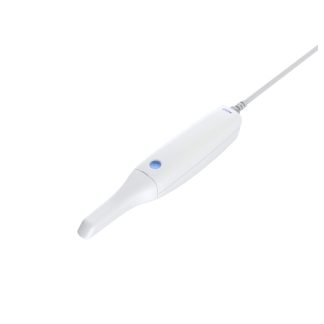
The Aoralscan Elite Wireless gives you the Elite’s intraoral scanning and built-in photogrammetry with a rechargeable battery and wireless connection, so you can move freely around the chair on everything from single crowns to full-arch implant cases.
Key features
No cable at the chair
A rechargeable lithium-ion battery and wireless connection let you scan from any position.
Intraoral photogrammetry (IPG)
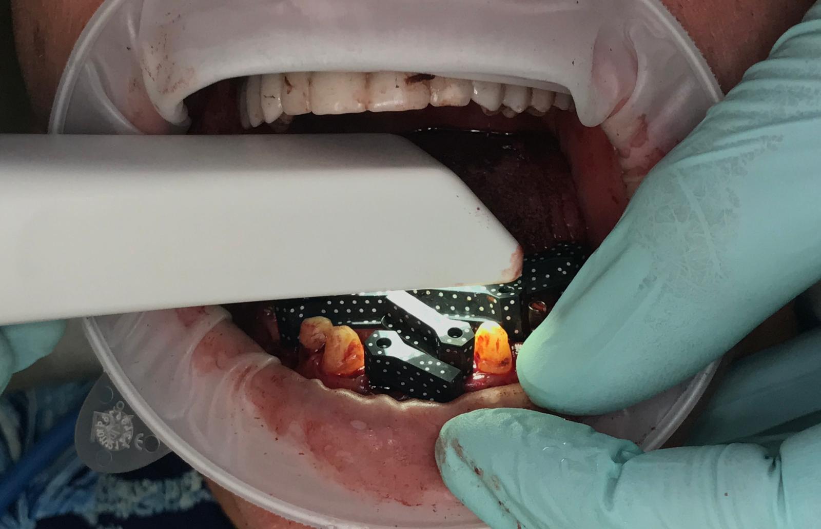
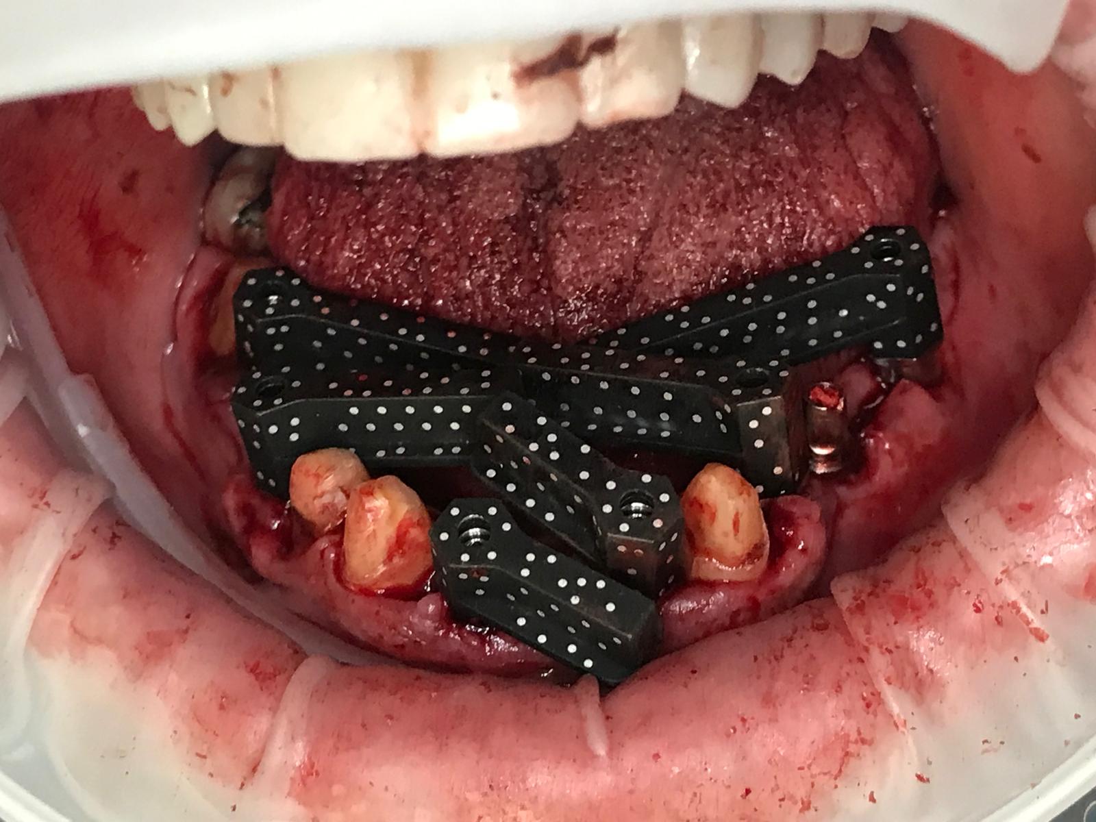
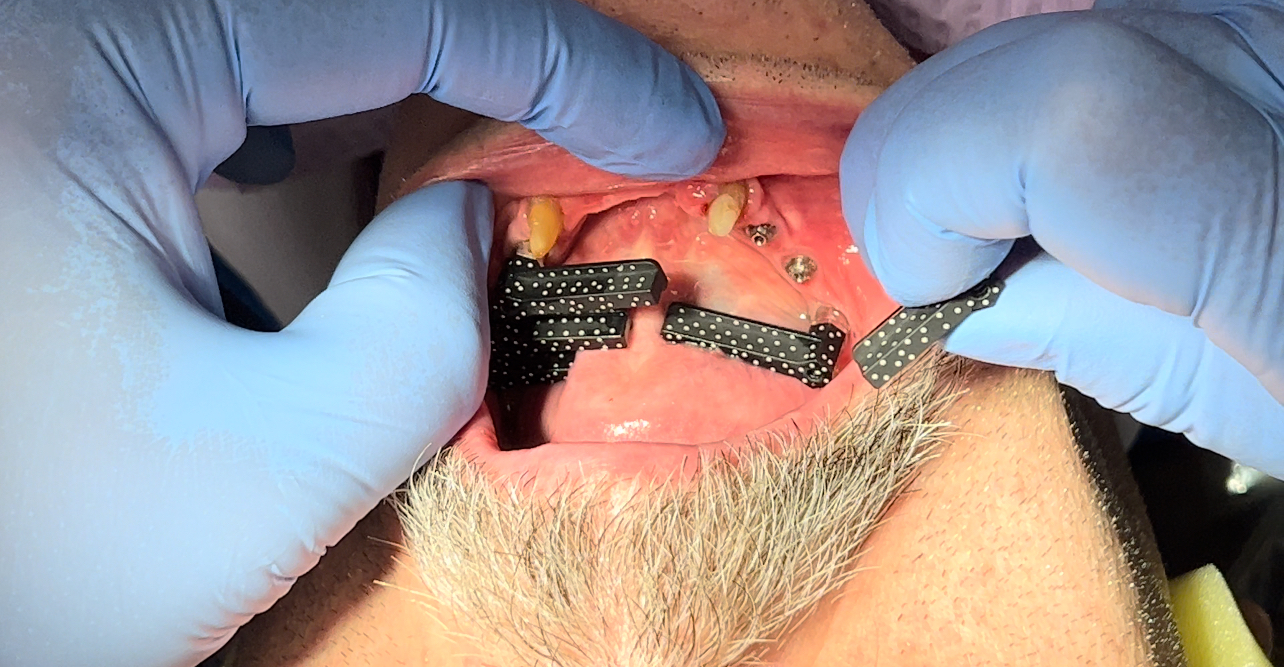
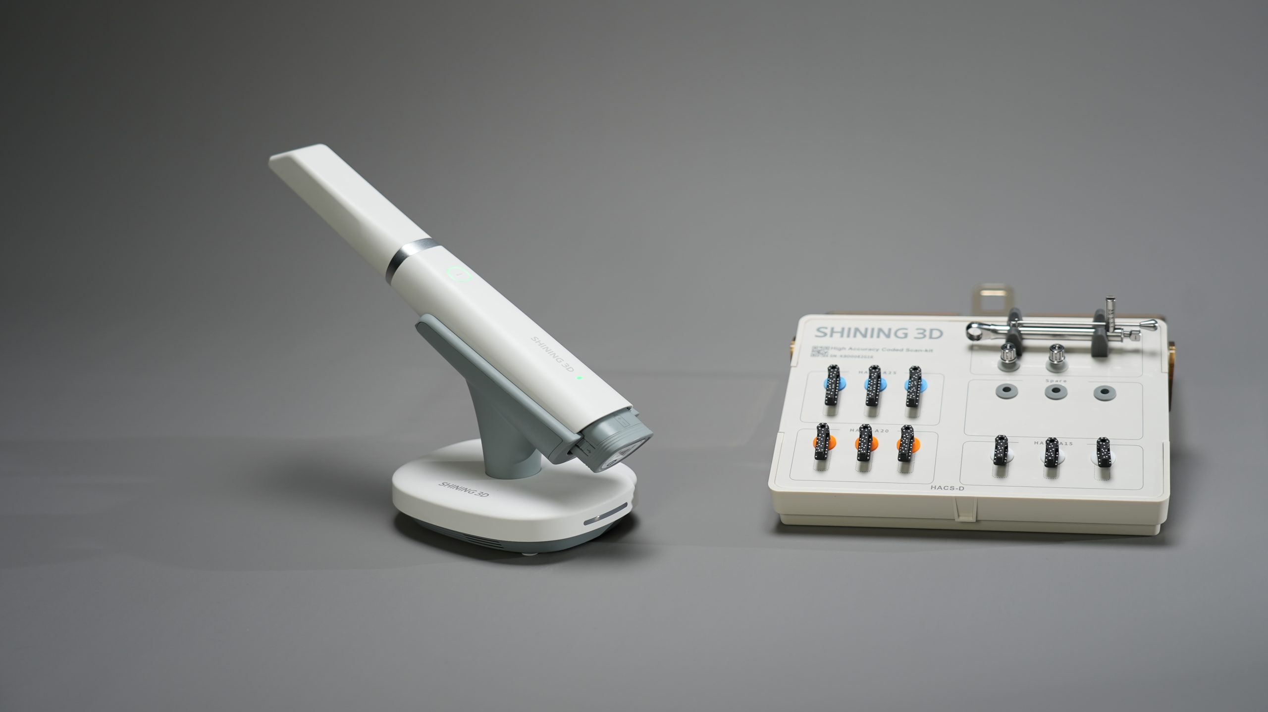
Capture full-arch implant positions with coded scan bodies and the large IPG tip, with 5 µm IPG precision, all in the same scanner you use every day.
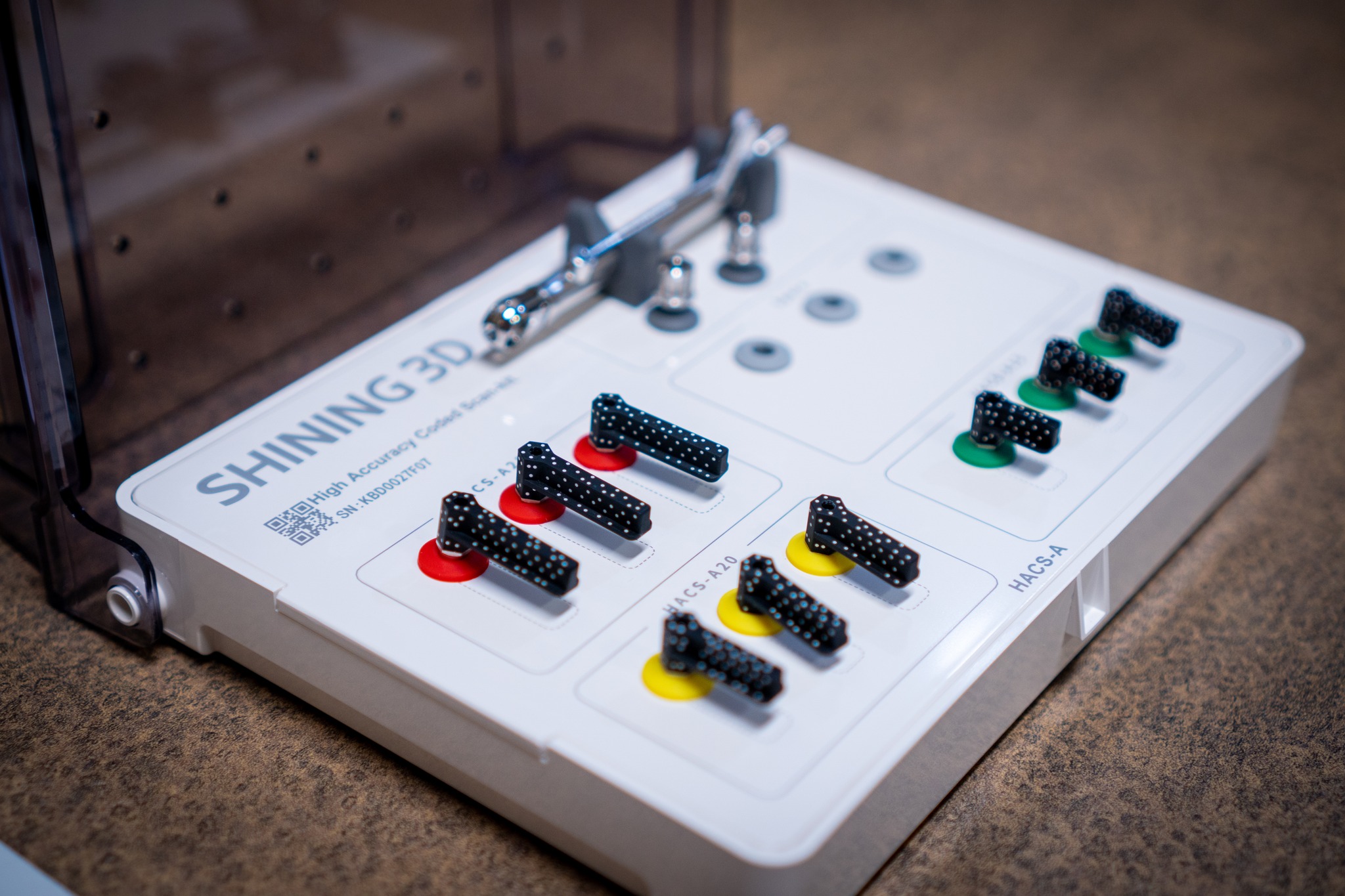
Coded scan bodies
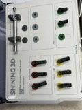
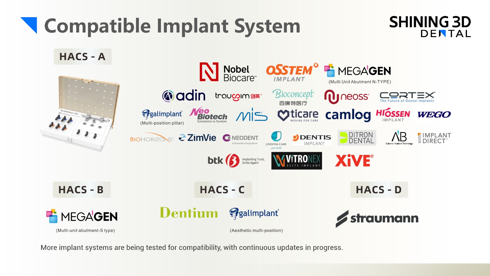
Shining 3D’s screw-detachable coded scan body kits fit more than 100 implant systems. Coded scan bodies work only with the Elite and Elite Wireless.
Three tip sizes
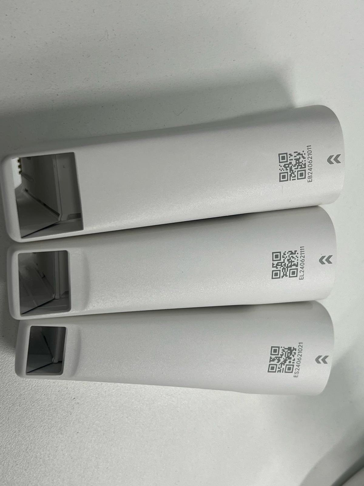
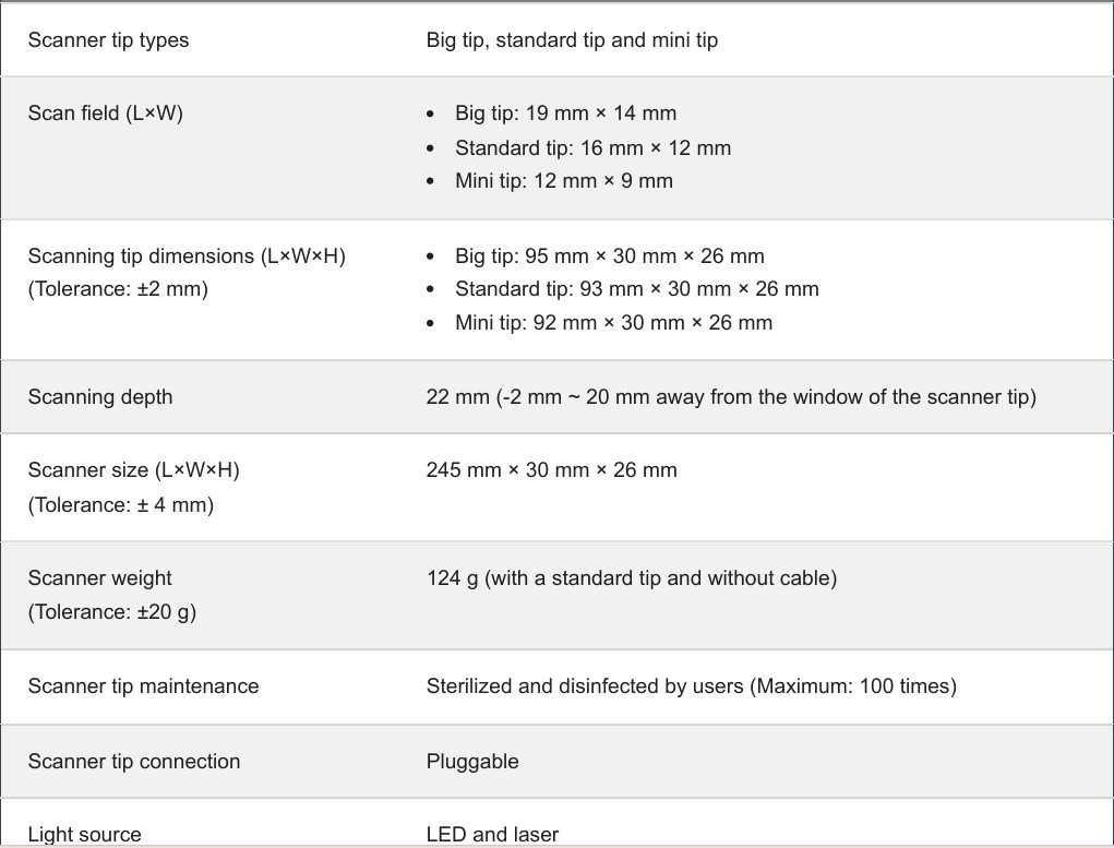
Large (IPG), standard and mini tips for full arches, everyday crown and bridge work, and tight posterior areas.
Aoralscan Elite Wireless Specifications
| Category | Aoralscan Elite Wireless |
|---|---|
| Weight | 194 g (with standard tip and battery) |
| Dimensions | 247 × 38 × 37 mm (with standard tip) |
| Connection | Wireless (battery powered) |
| Battery | 3250 mAh, 3.6 V lithium-ion; at least 80% capacity after 400 cycles |
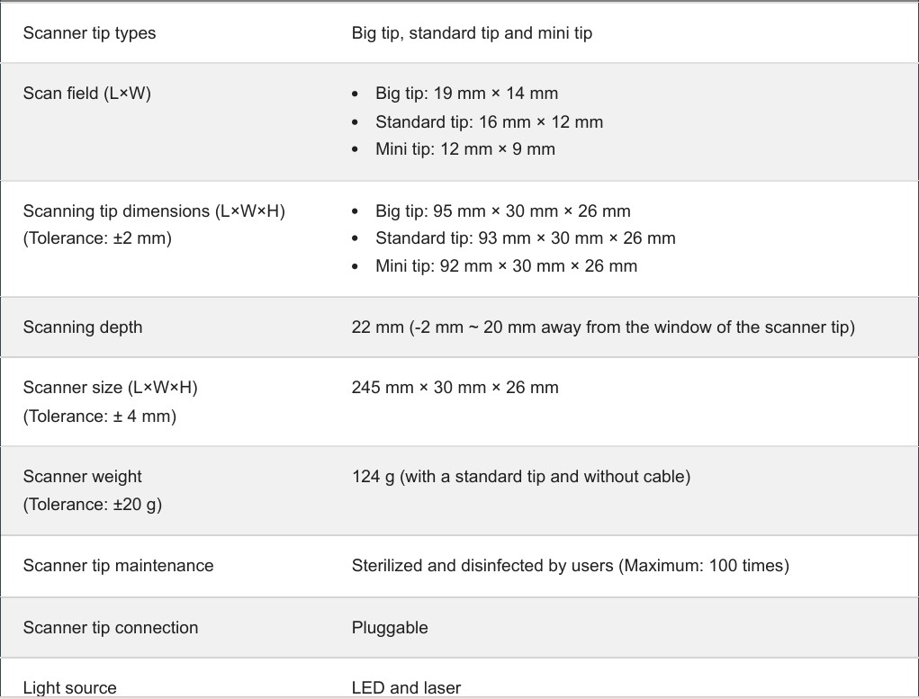
| Scan fields | Large (IPG) 19 × 14 mm, standard 16 × 12 mm, mini 12 × 9 mm |
| Scan depth | 22 mm |
| Cradle | 119 × 93 × 101 mm, 340 g |
| Output formats | STL, OBJ, PLY |
| Photogrammetry | Built-in IPG with Shining 3D coded scan bodies |
Specifications from Shining 3D.
Package details & unboxing
SHINING 3D NEW Elite Intraoral Scanner – WIRELESS – April 2025
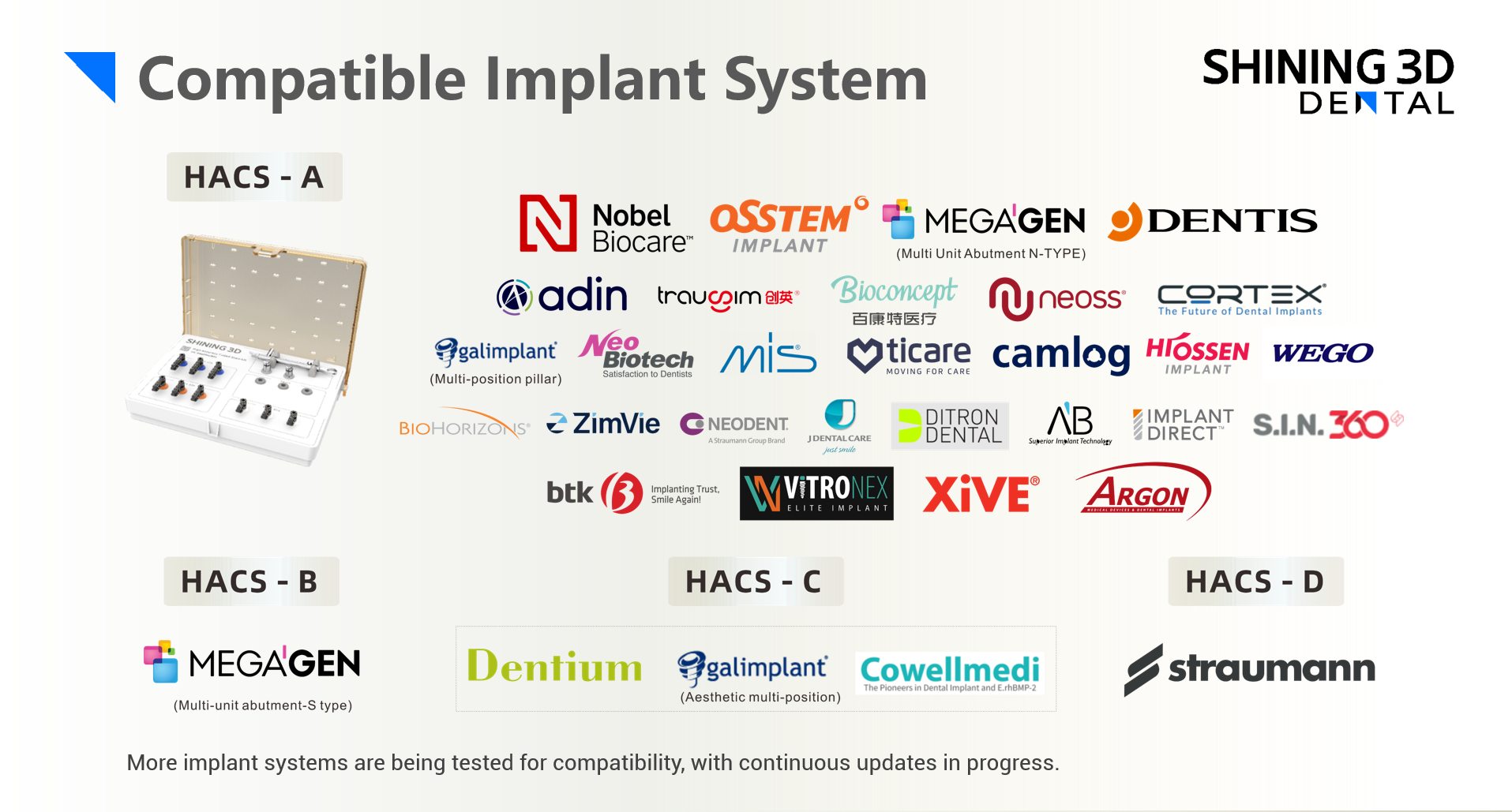
Included HACS-A Kit – other Kits Available here:
Unboxing video:
Why buy your Aoralscan Elite Wireless from CAD-Ray?
- Training and support from CAD-Ray, one of the largest independent CAD/CAM dealers in the U.S.
- CAD-Ray sells the full Shining 3D line, including tips, scan bodies and accessories, so your whole Shining workflow is supported in one place.
Aoralscan Elite Wireless FAQ
What is intraoral photogrammetry?
Photogrammetry captures the exact 3D position of multiple implants using coded scan bodies. It’s built into the Elite, so you don’t need a separate photogrammetry device for full-arch implant cases.
Which scan bodies does it use?
Shining 3D’s high accuracy coded scan bodies. The screw-detachable kits A–G cover more than 100 implant systems; see our Shining 3D accessories page for which kit fits your system.
How is the Elite Wireless different from the wired Elite?
It adds a rechargeable 3250 mAh battery and wireless connection, and weighs 194 g with tip and battery. The scanning, photogrammetry and coded scan body workflow are the same.
Clinical cases & videos
Shining’s Elite Scanner combines incredibly efficient intra-oral imaging with photogrammetry for perfectly capturing implant locations.
Here is a quick introductory video of how the process works and the subsequent cases provide a lot more detail and information
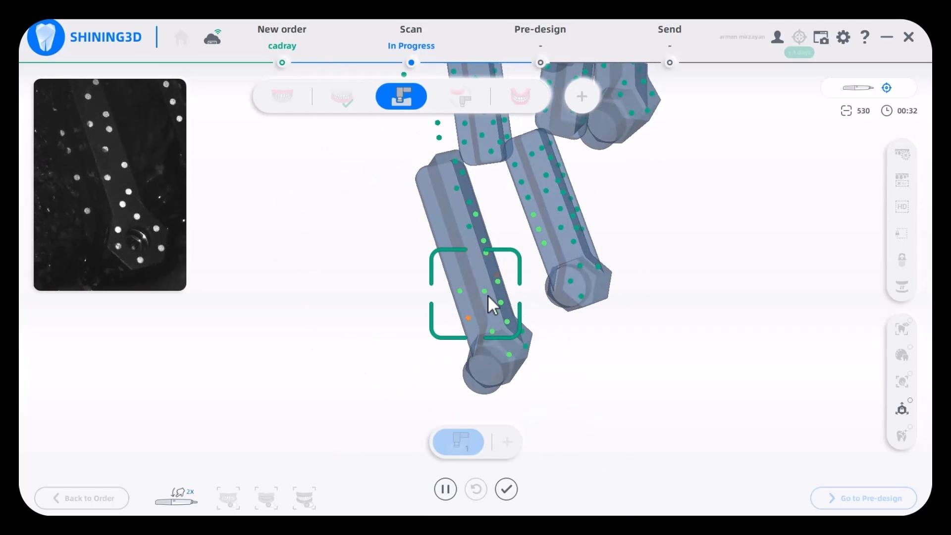
Case 1
In this video we detail how the process works and how you capture everything you need for your full arches with one scanner.
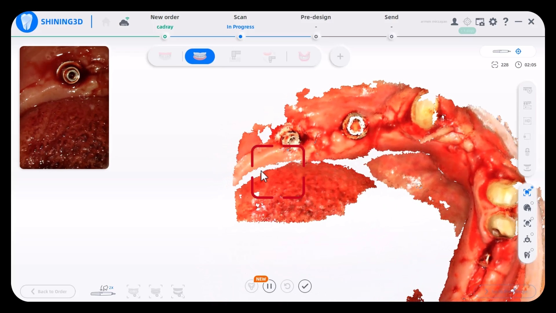
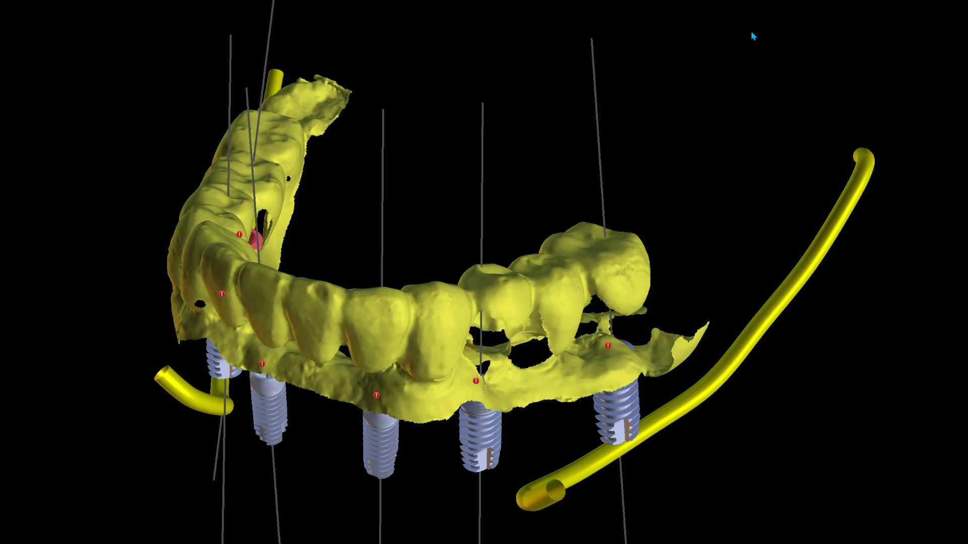
Precision, Trueness, and Accuracy
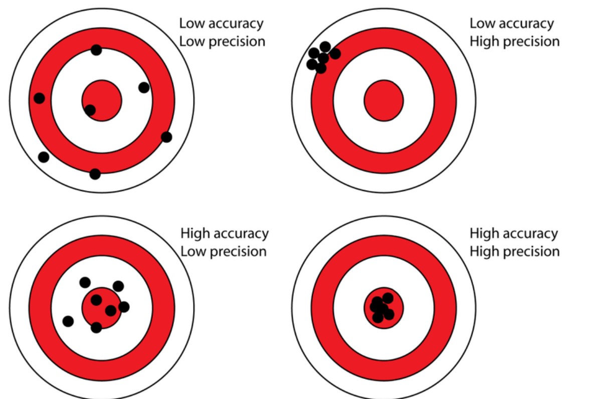
A common property that is oftentimes used to describe how well scanners perform is how accurate, precise, or true it is. A lot of people use this incorrectly to describe intraoral scanners and it is utilized in poor judgment because there are too many variables in the input of the equation to ascertain any conclusion from the output file. But these characteristics CAN be used to study photogrammtery.
In this chart, the bullseye represents the true value (true location of the object), while black dots represent measurements (the estimated 3D locations of the object based on the 2D images) from this source. To measure the precision, we took 3 random arrangements of the dominos and found the output files to be identical. To measure its accuracy and trueness, we related it to a desktop scan of the same model
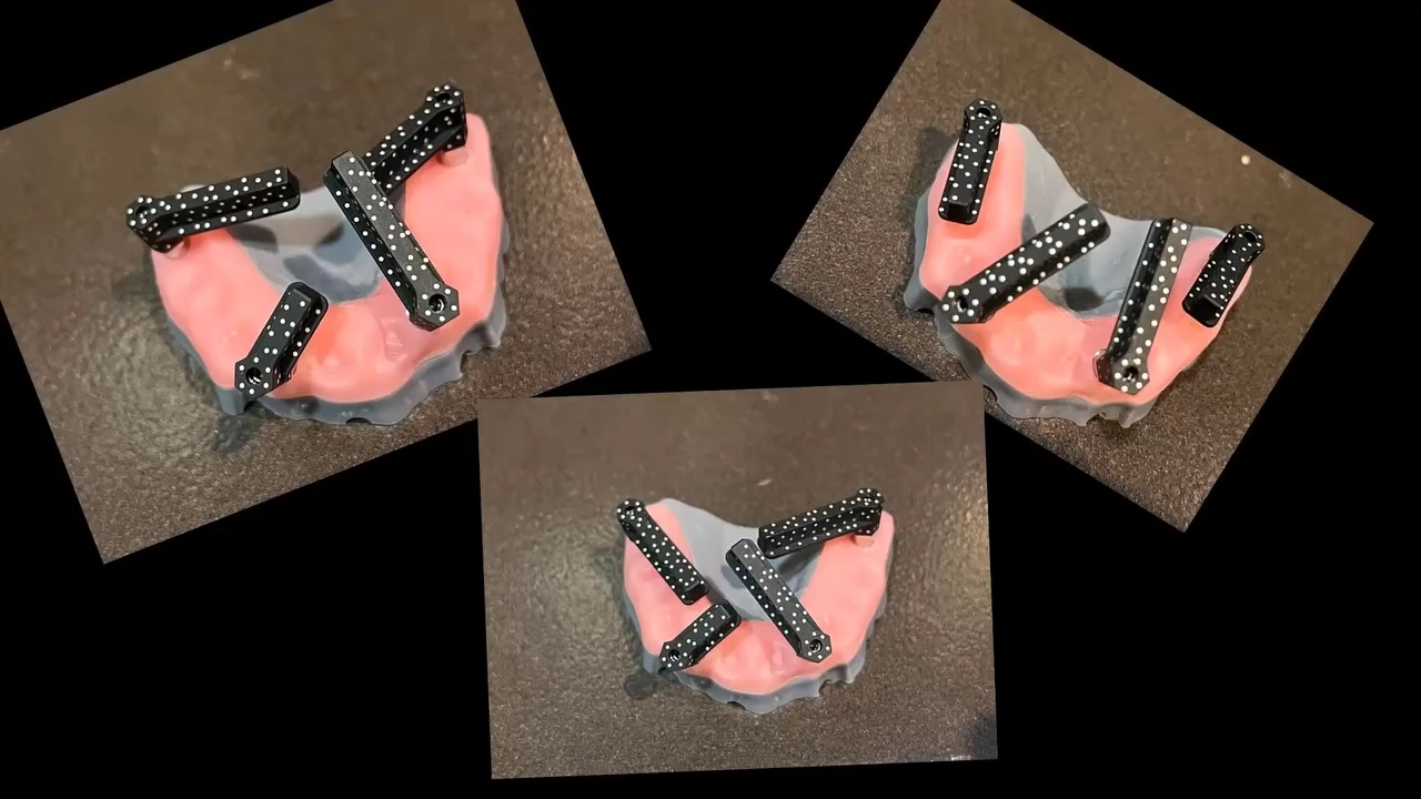
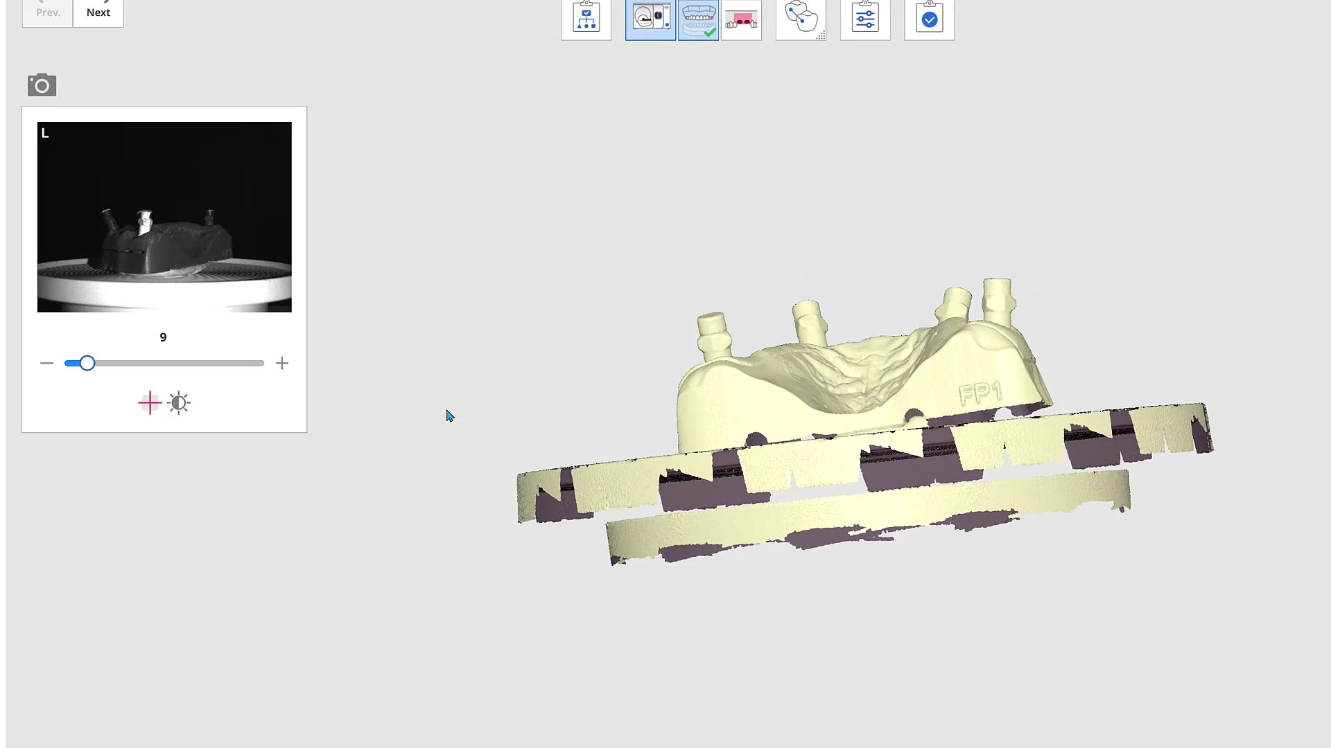
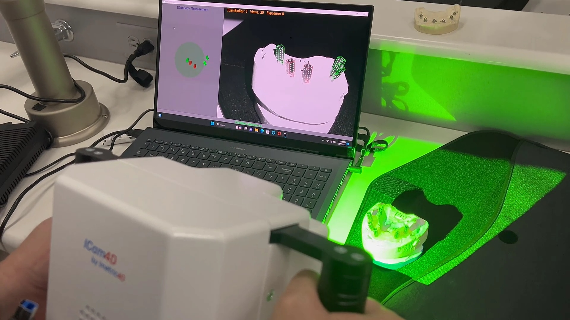
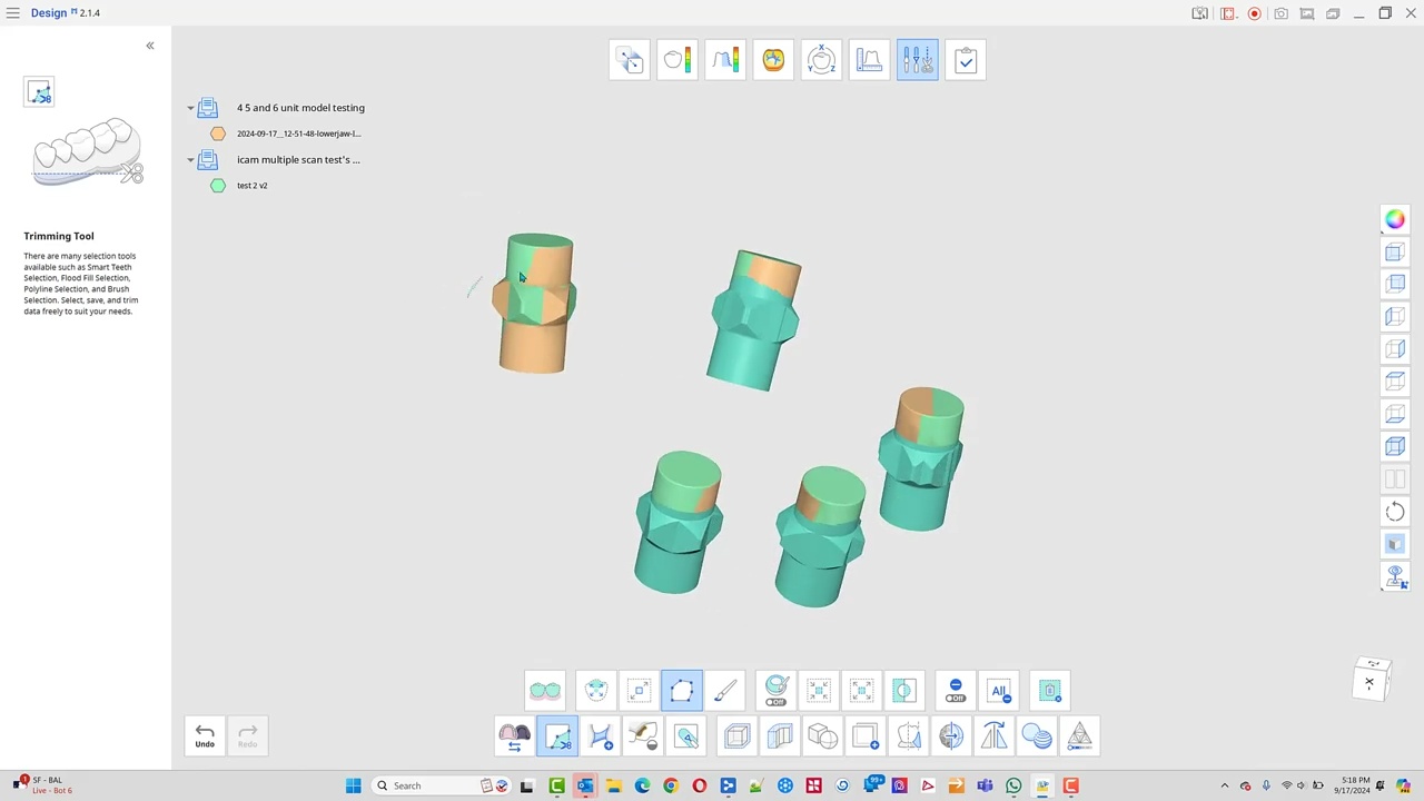
you can download these models and test them out for yourself: compare elite to icam and desktop scan
When is image capture complete?
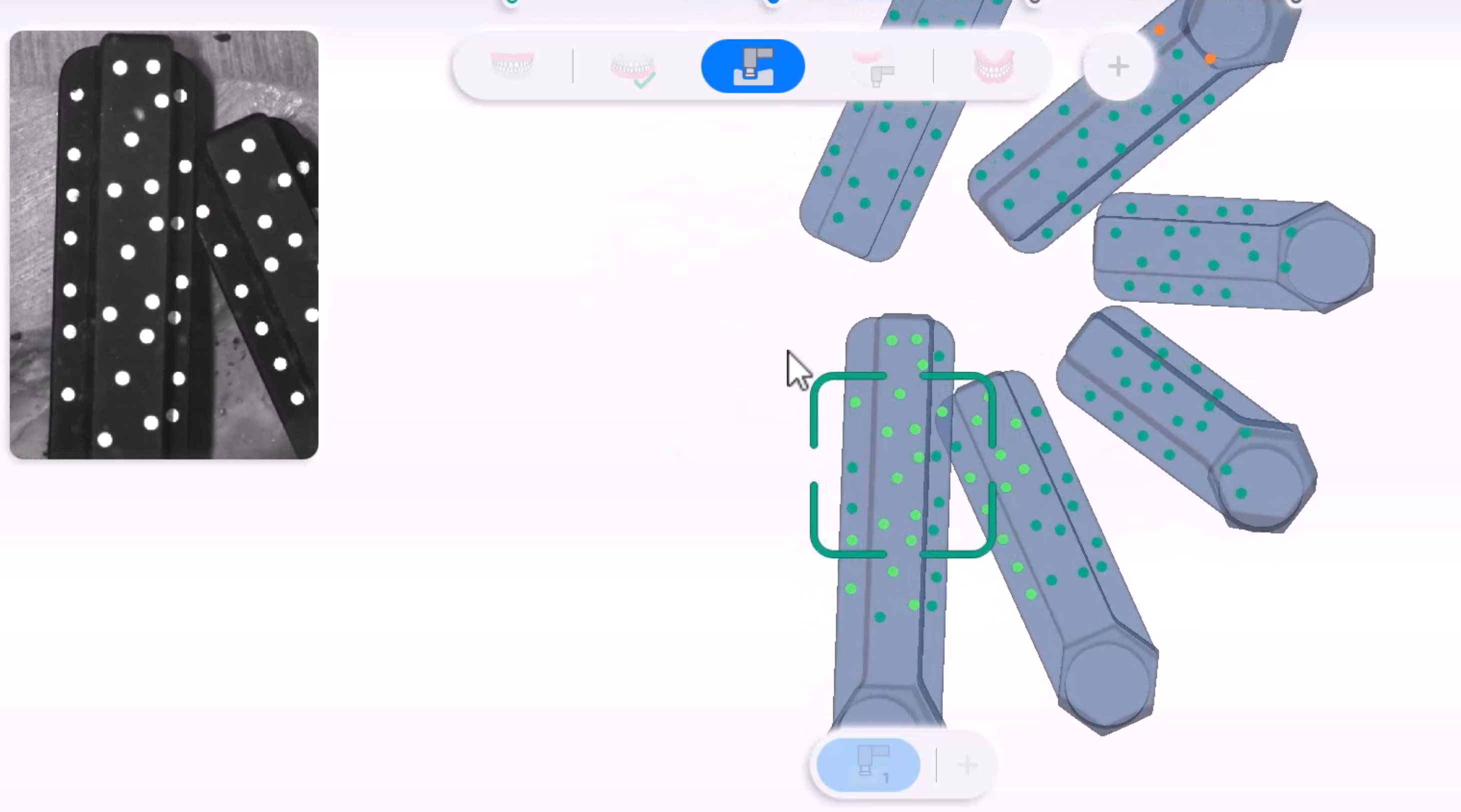
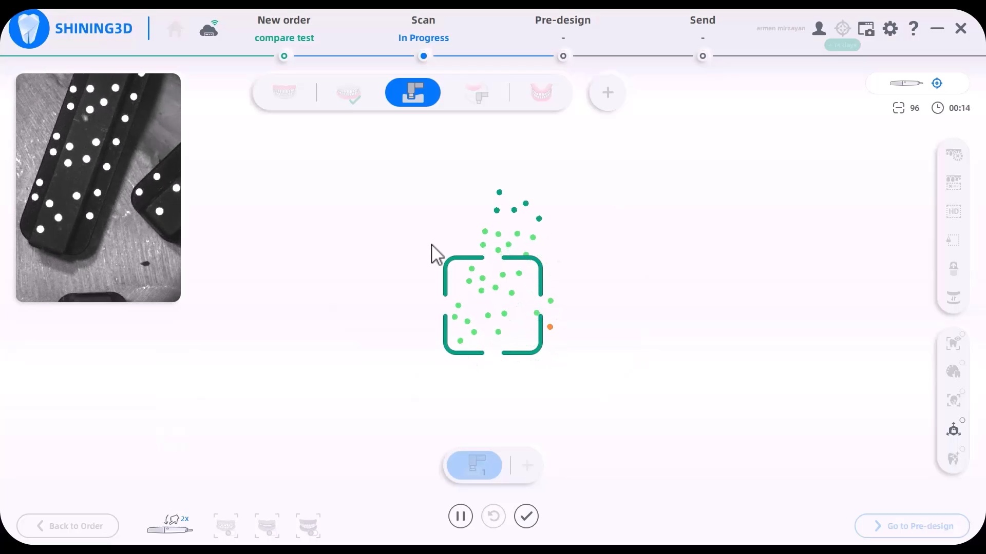
A frequent question we field is how does an operator know how much of the dominos / scanflags to capture. The manufacturer recommends a specific scan path and to completely capture the suprastructure. Our internal testing shows that there is no difference between the results as long as you allow the software to spit out the scanflag geometry while you are scanning. See this video for an explanation
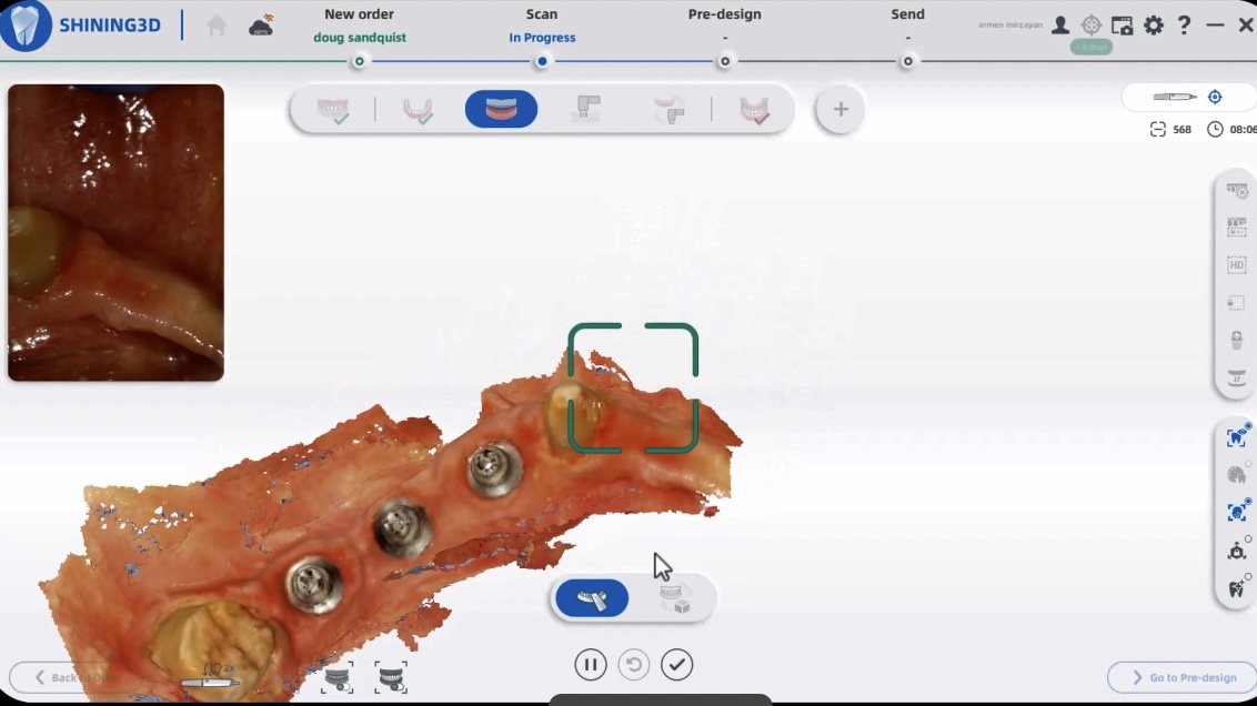
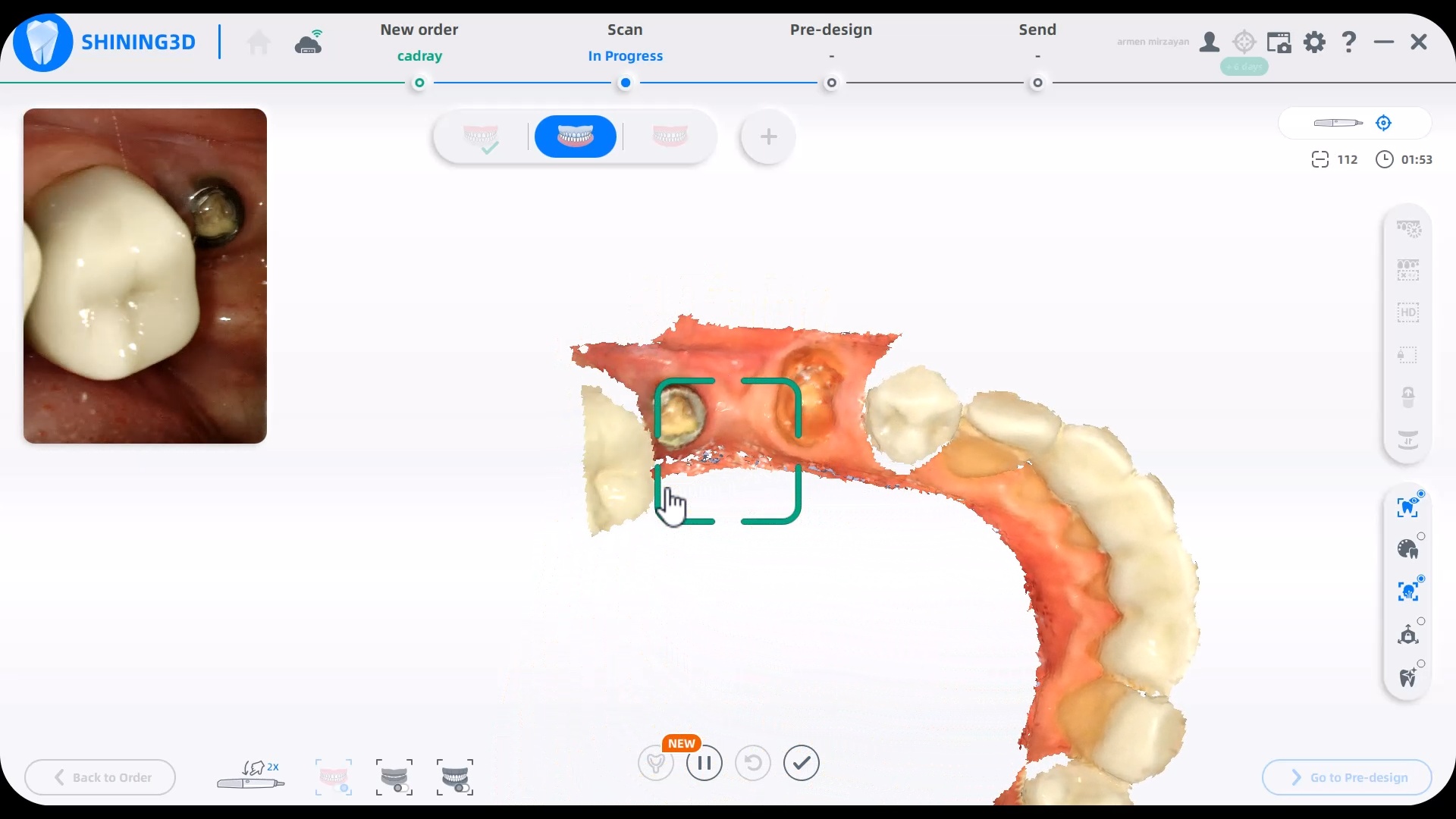
How Well does the Intraoral Scanner Work on Soft and Hard Tissue?
Case 2
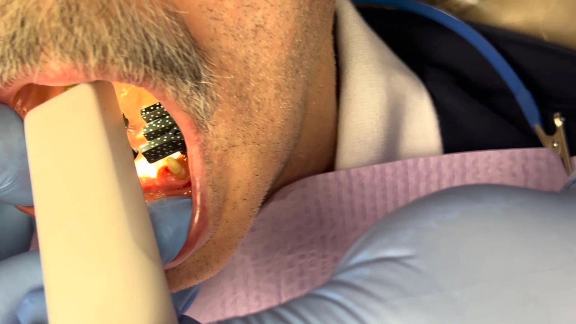
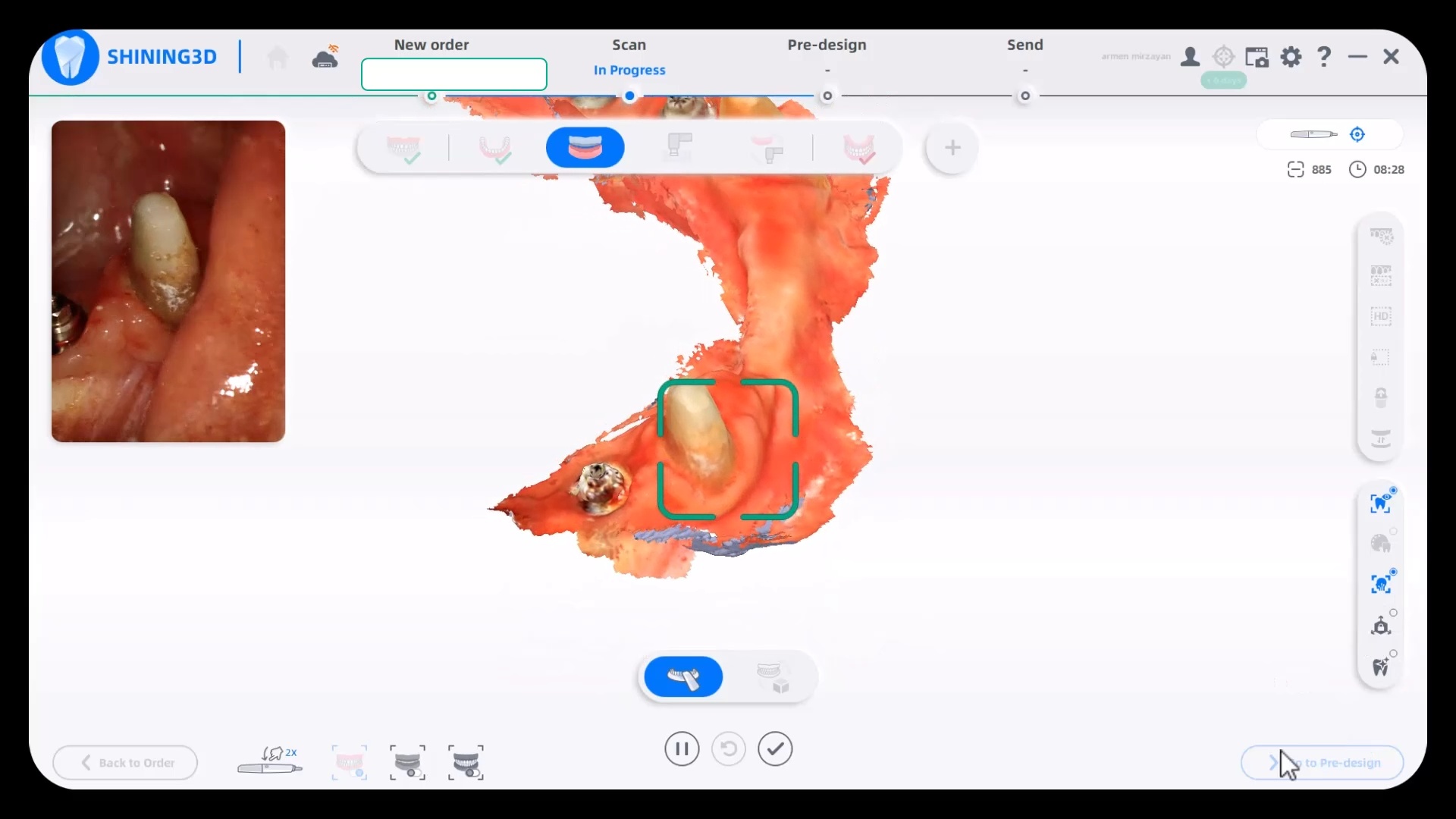
The first video in this series shows a quick overview of a multi unit case and the subsequent videos goes into more detail
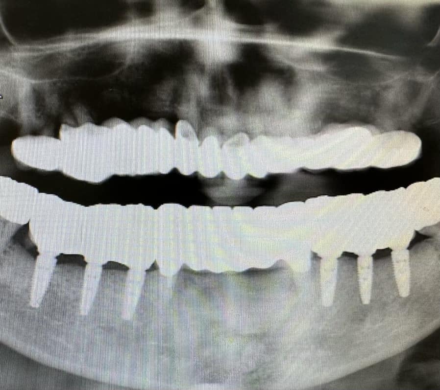
Radiograph of the delivered case
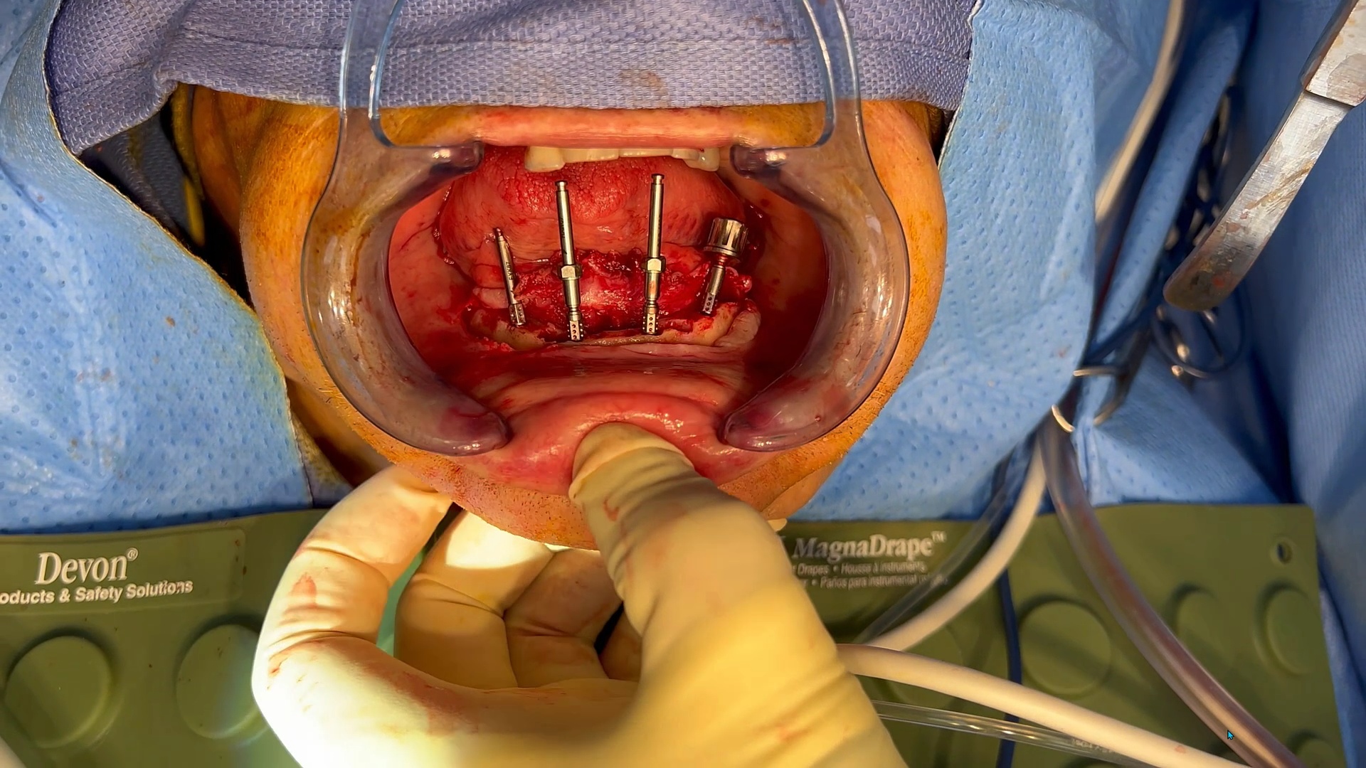
Case 3 – Managing and immediate case with bone reductio
This video shows how to manage immediate conversion cases and compares and IPG with an extra-oral Photogrammetry Machine
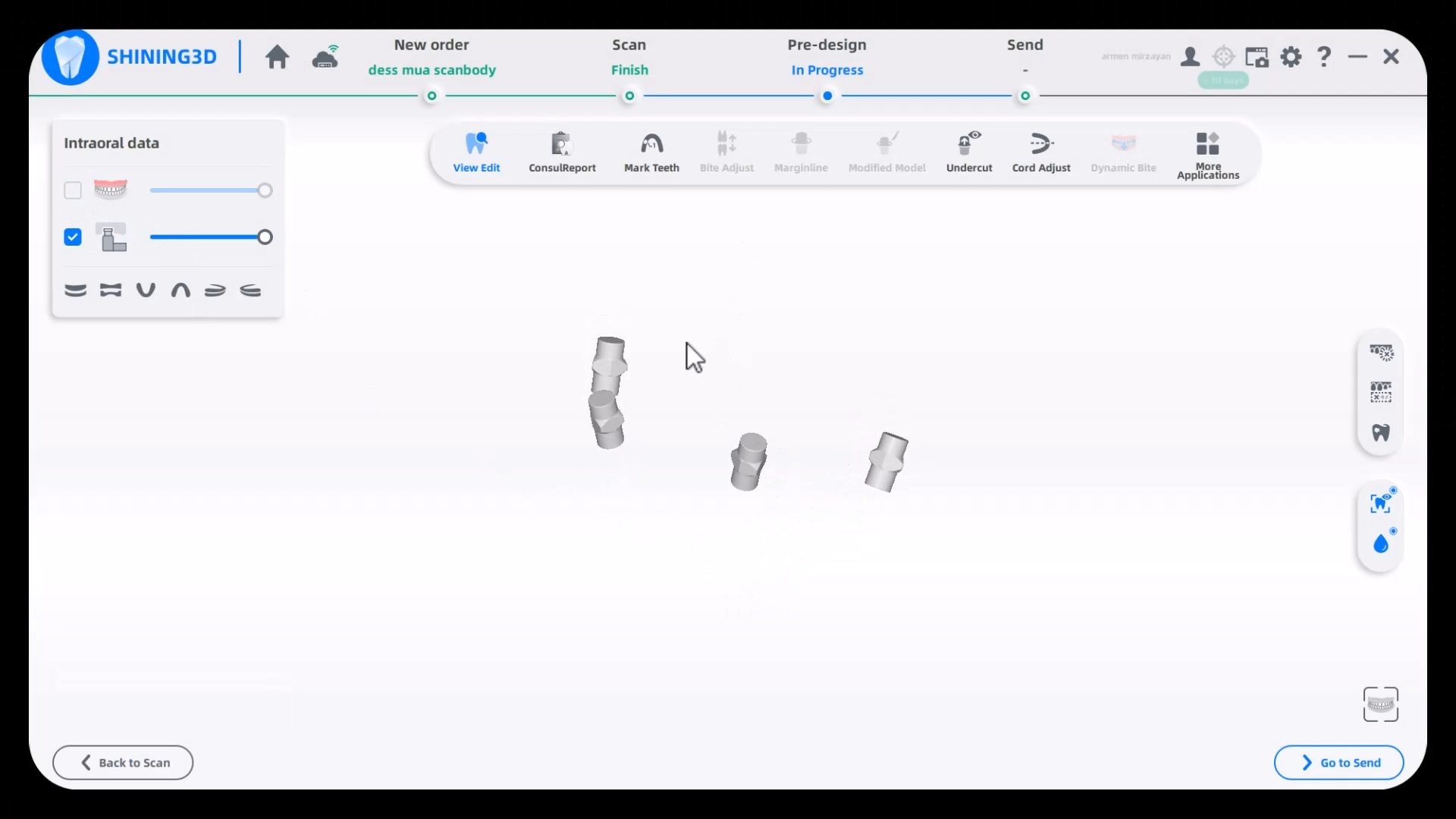
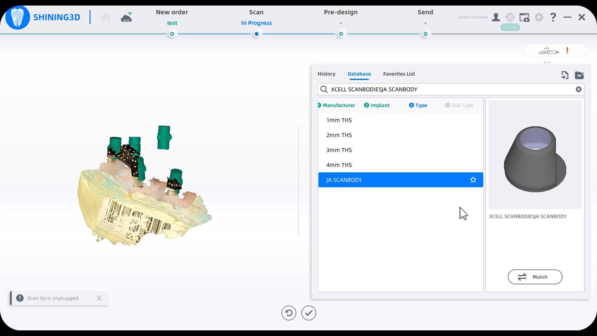
How to load libraries into Shining 3D Elite Intra-Oral Photogrammetry Software
This should be done by an experienced person. Here we load a dess MUA library into the equation in the first video. In the second video we import Jonathan Abeneim’s Xcell libraries, followed by Danny Domingue Vortex library import
Scanner Tip Dimensions

Reviews
There are no reviews yet.