


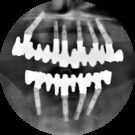


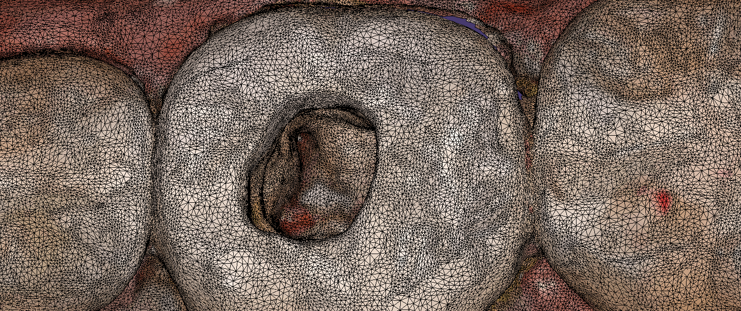
Imaging depth and red tissue or blue can be complicated for all intra-oral scanners. In this demonstration case, we show how the Medit i500 performs scanning inside the canal space of a lower first molar after a pulpectomy. The dry canals are readily picked up by the scanner at a focal length of 21 mm. The bleeding from the distal canal space makes it a bit more challenging to capture.
Medit i500 uses two cameras that operate at different filter levels of color spectrum. The camera was changed from blue (great at capturing tooth structure) to white camera, which is better at capturing red (soft tissue and bleeding)
