


Implant Scanbody Imaging with Medit i500
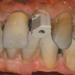 There is a great feature in the Medit Link software that allows you to scan implant scan bodies and instantly locate the fixture location, timing, and angle. What is great about the process is that you can image the edentulous area before the scanbody is placed onto the fixture. When you go back and scan the scan body, it only images what is “different” between the two catalogs and you get awesome models like this:
Here’s how it is done: when you image the adjacent teeth, you want unimpeded access to all the information below the height of contours of the adjacent teeth. This is impossible to accomplish if you have the scan body in place, as you won’t be able to get around it.
So you just scan the arch, and then scan the same arch with the fixture in place. You will see in this video how there is a peri-osteal flap raised and lots of “activity” around the scanbody, but the software only picks on the Peek ScanBody, and ignores all everything around the area that is already plotted. Watch the video and you will understand very quickly how important this feature can be.
[videopress rqtTMT8Y]Download the STL
Download the OBJ
Download the PLY
