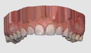


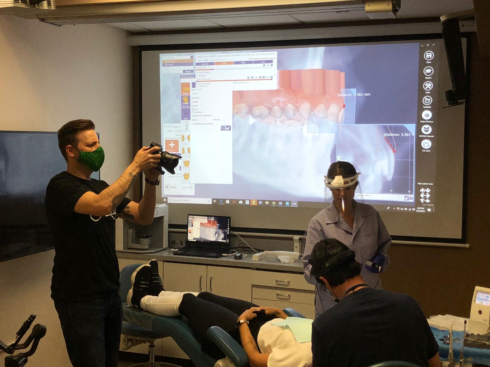
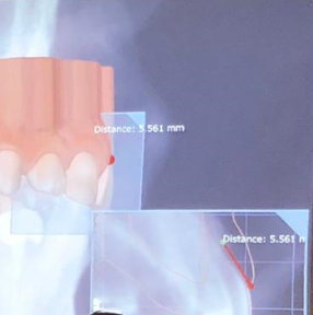
 A Medit i500 scan was taken and merged with a CT scan and the distance to bone was measured (5.5 mm’s) giving enough space for soft tissue contouring with a diode laser. The 3D X-ray must be captured with cotton rolls placed in the vestibule so that you can differentiate between the gingiva and the lip. Ideally you should not close the base of the digital model by the intra-oral scanner as it can make the merge of the data sets more difficult than it needs to be.
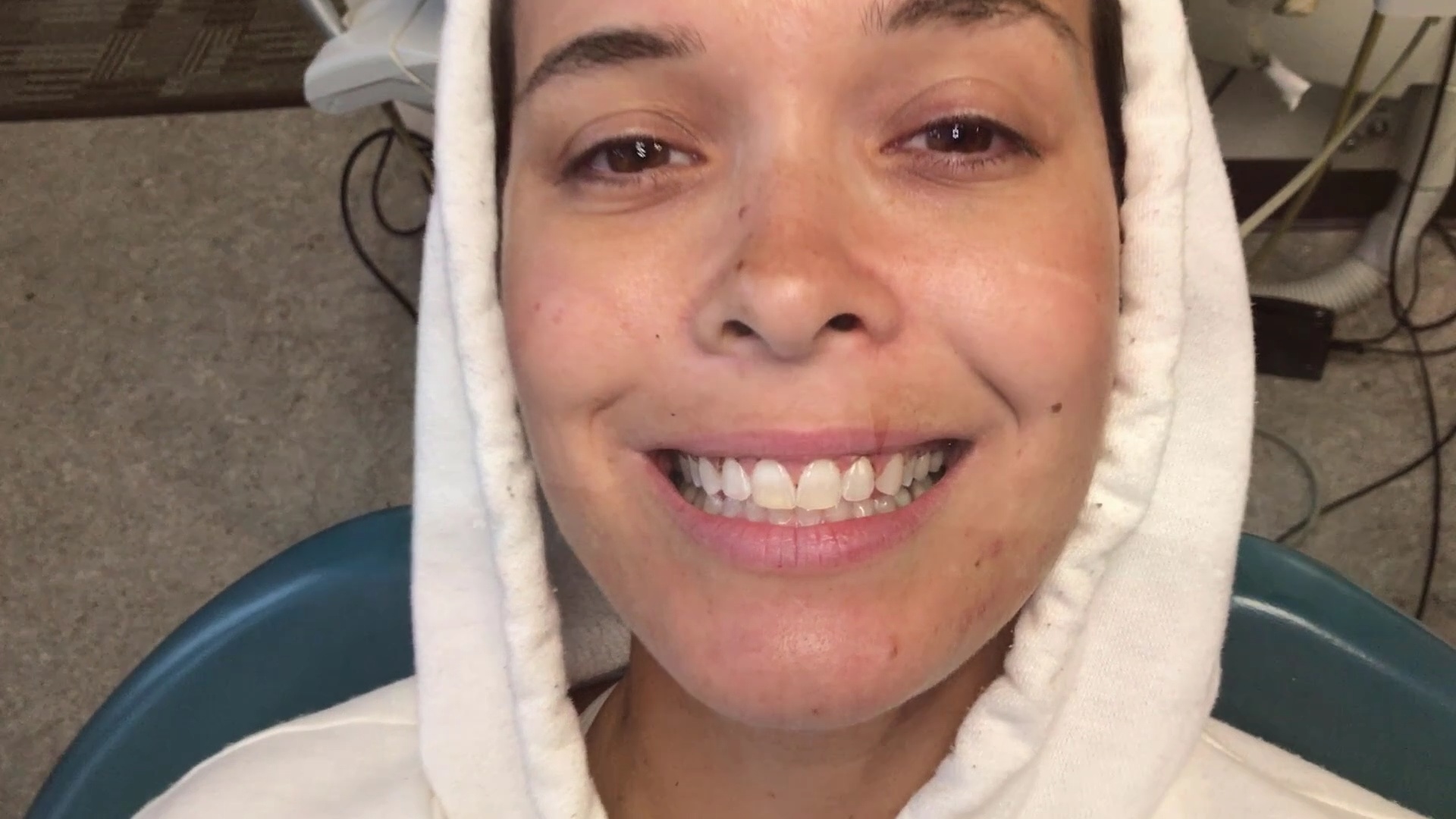
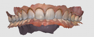
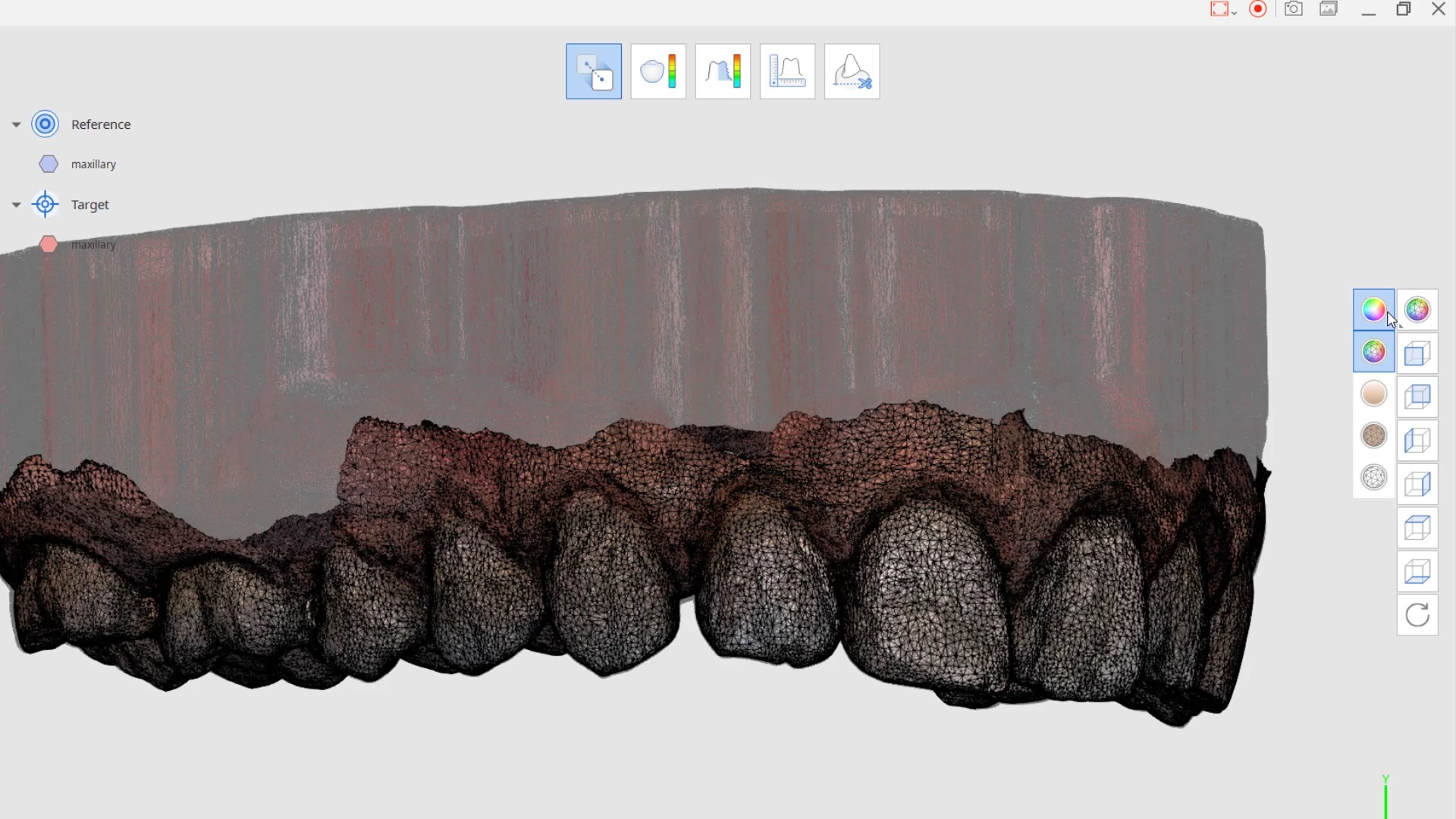
Comparison of Pre-Op Scan and Post Gingivectomy Scan with Medit i500