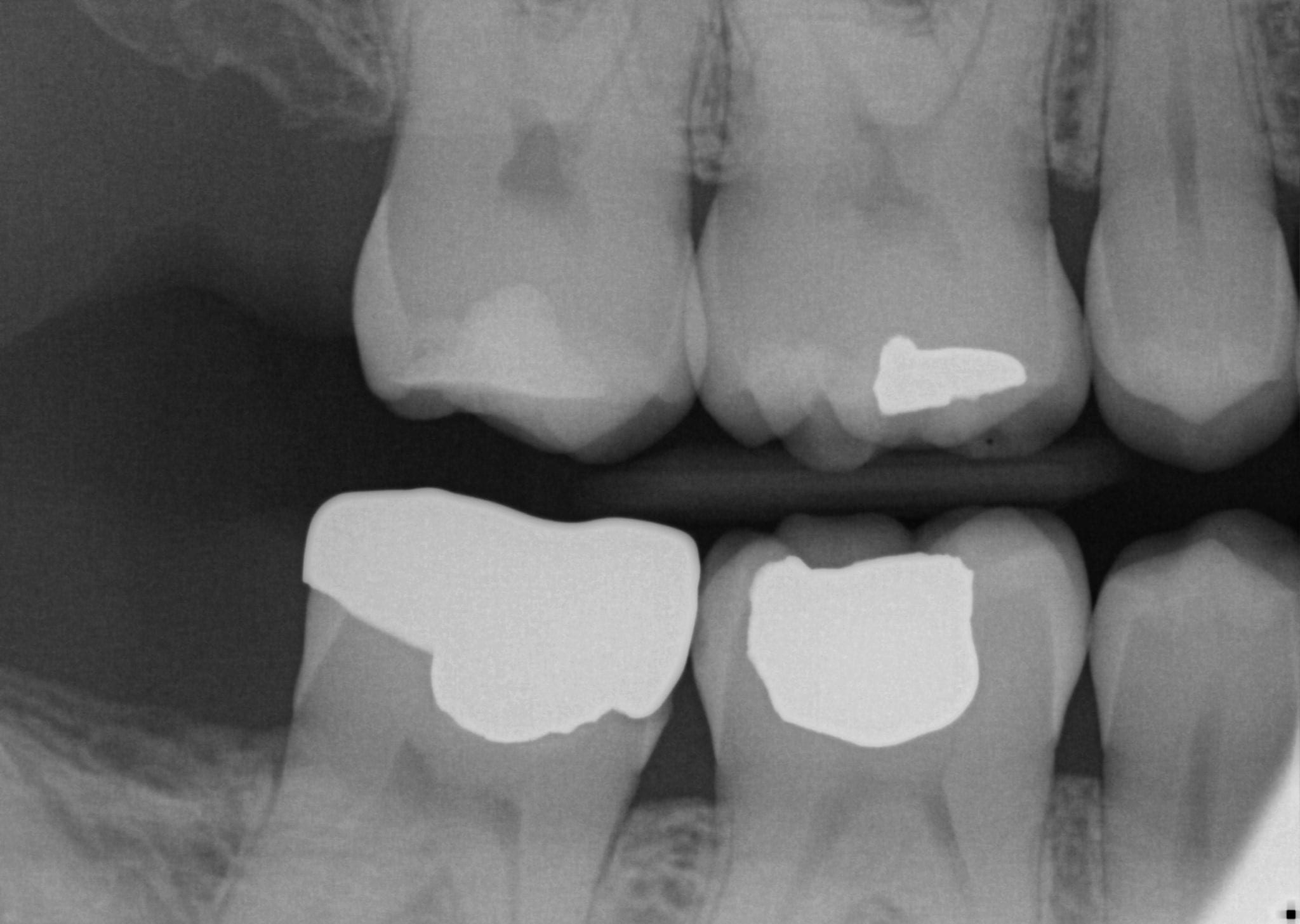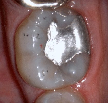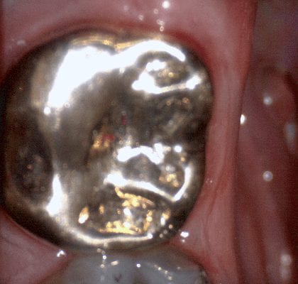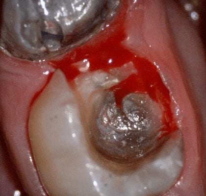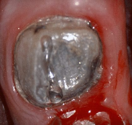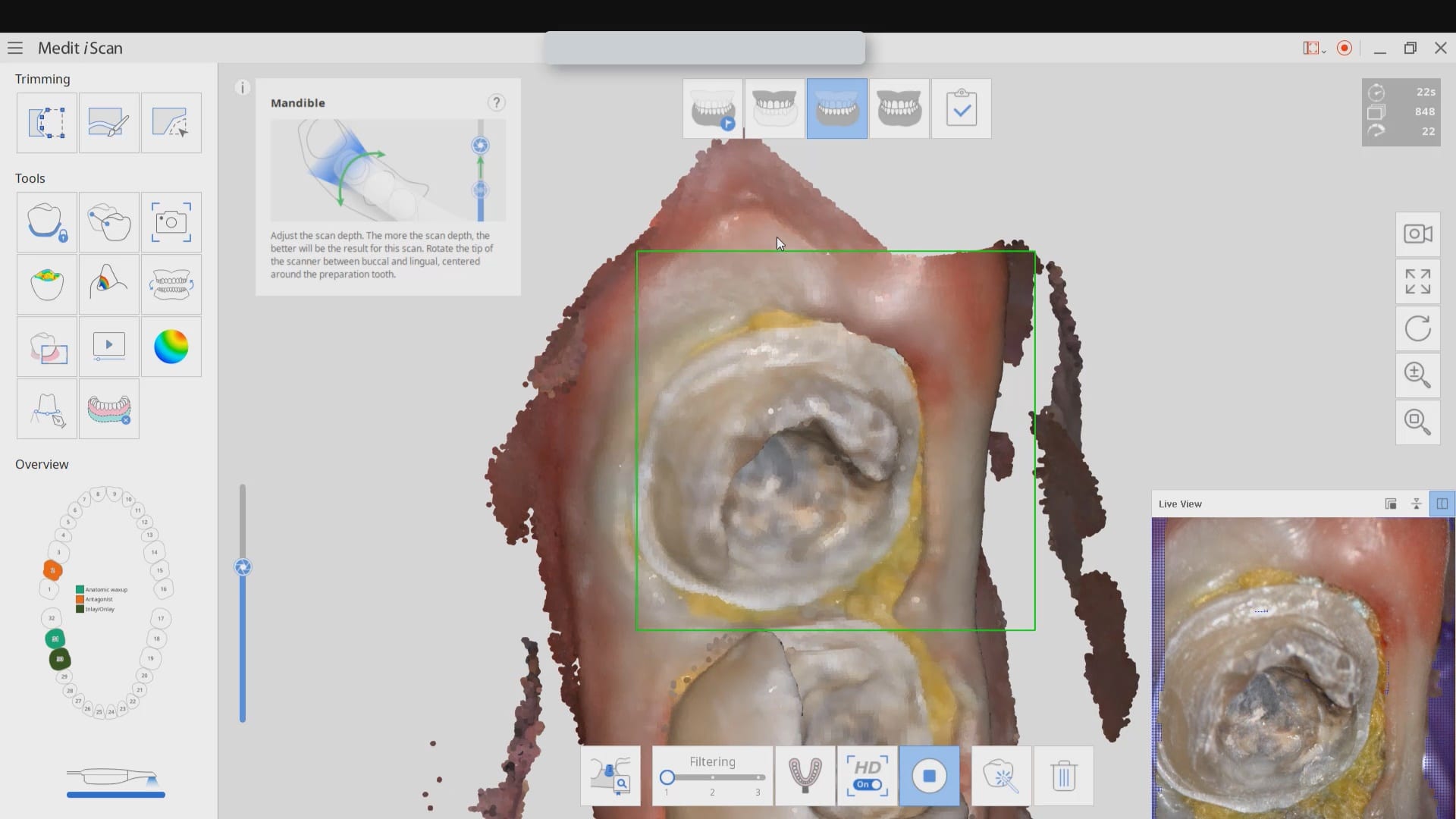


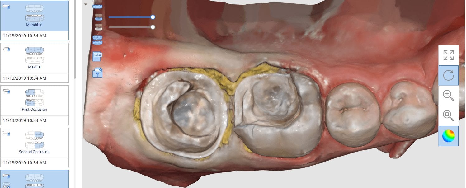
In this video we demonstrate some of the most common problems associated with second molar impressions. Usually the distal margins can be blurred by the presence of hemorrhaging or soft tissue. In this particular case, we use the tip of the camera to displace the tissue and digitall correct an area for better accuracy. The isolite systemisolite systemisolite system does a great job controlling the tongue and the lips and with proper retraction you can image a quadrant in very little time
The second most commong problem is a change in the vertical dimension before and after we prepare the last vertical stop in the arch. We advocate that you take two bites; one before preparing the distal extension and one after you prepare it. Comparing the two buccal bites will let you know if you will have issue with your vertical dimension and allow you to reduce the post op adjustments