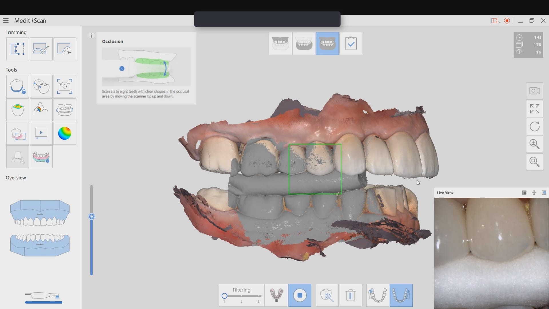


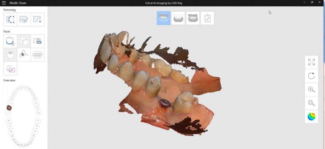
This case shows a full arch imaging where the model starts to go “off track”. To keep the explanation simple, the further away you get from multiple planes / heights of immobile structures, and the more you image in flat areas, you can inadvertently introduce errors in your models. You can see a sample case here in the video.
[videopress zqRzFmTj at=”8″ permalink=”false” hd=”true” autoplay=”true”]There is a very simple solution, as you can see in this video. It entails consistently moving back to reliable landmarks to stitch new information to exisitng correctly. If you notice how we start imaging on the occlusal of the premolars and the molars, then we roll to the facial of the molars and the back around to the palatal of the molars. Then we sweep over soft tissue on the palate. But we don’t continue in that direction. We immediately return to the molar area so the software has landmarks it can recognize.
We then move the camera forward, image the palatal of the premolars, and then sweep back across the palatal midline. We repeat this back and forth movement to maintain a proper path for the software to recognize landmarks that do not move, in the equation. With this technique, you can scan a whole upper arch in just a couple of minutes and capture great details of the dentition and the soft tissue on the palate
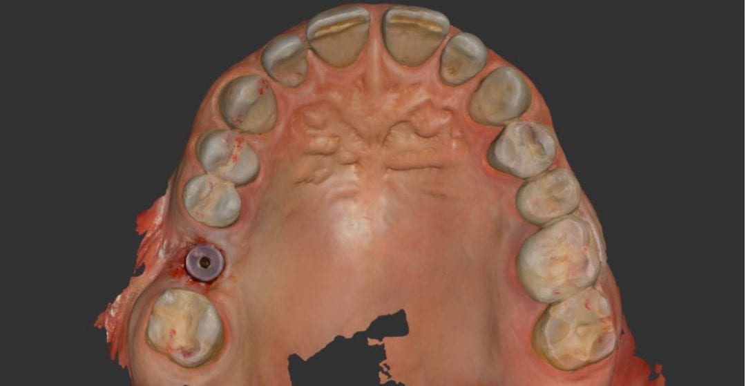
[videopress VVEbeoWs permalink=”false” hd=”true”]Here, you can see how the software rendered a perfectly accurate and detailed upper arch with the palatal vault captured without any errors.