Posted on Leave a comment

“Veersing” Effect – How Intra-Oral Scanners Introduce Errors in Edentulous Jaws and Flat Planes

In the world of digital dental scanning, precision and accuracy are paramount. To shed light on the potential pitfalls and challenges faced by dental professionals, Dr. Armen Mirzayan of CAD-Ray has put together an eye-opening video. In this demonstration, Dr. Mirzayan explores the intriguing concept of The “Veersing” Effect and how it can lead to scanning errors when using digital dental scanners. We’ll delve into the video to understand the nuances of these errors, using examples from some of the leading brands we sell, including the Trios 5, Dexis 3800, and Medit 700. Each of these devices possesses its own unique features and capabilities, making them essential tools in modern dentistry.

Understanding Scanning Errors:

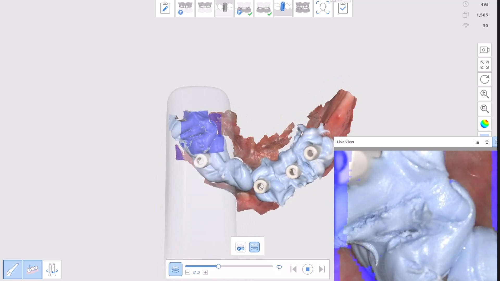
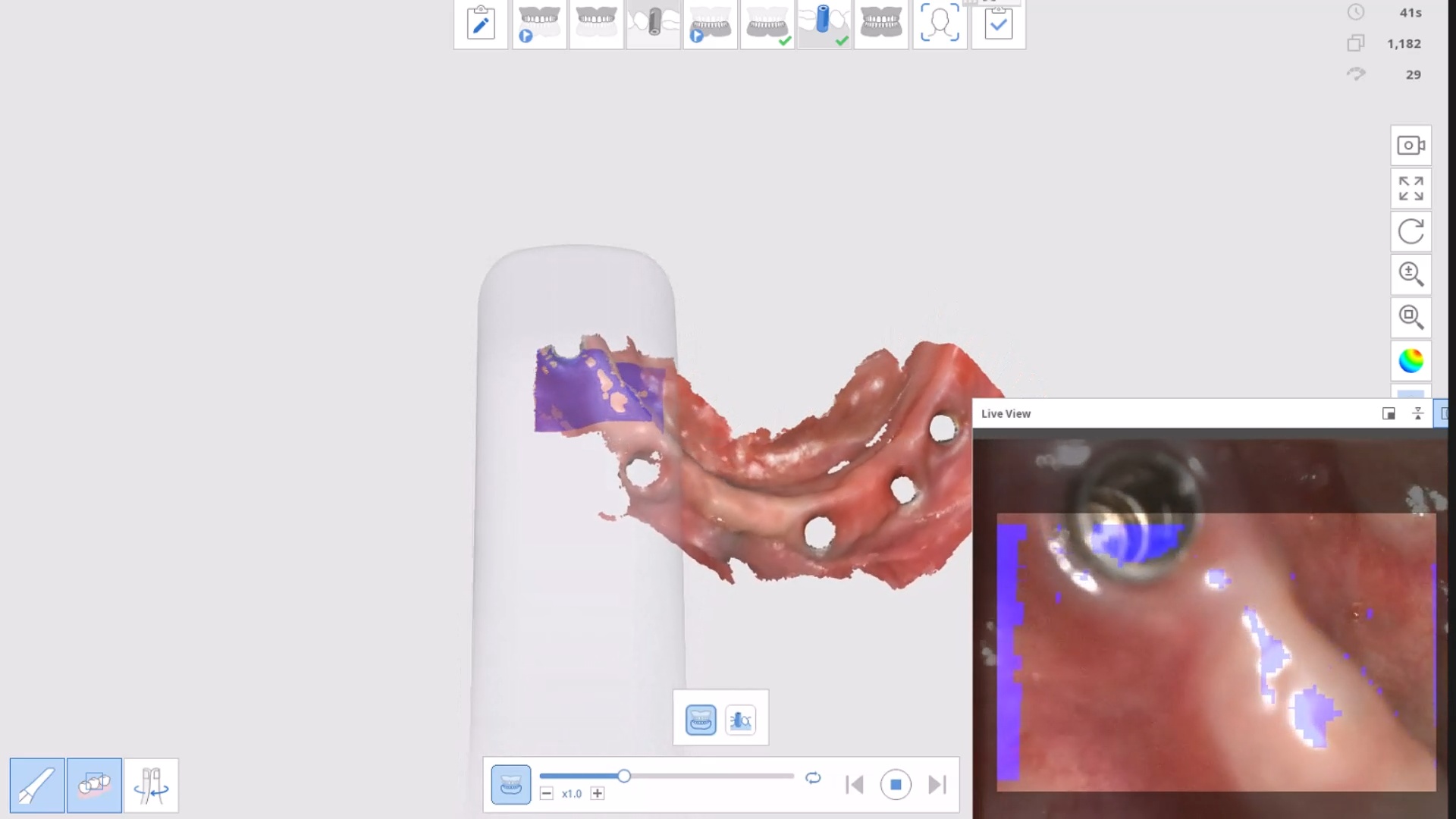
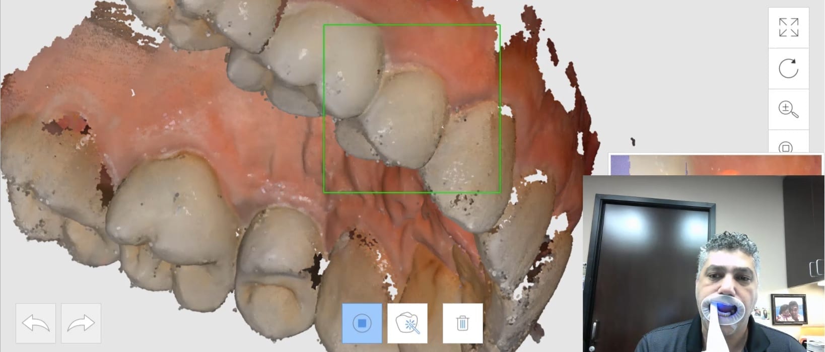
Scanning errors can have significant consequences in the dental industry. From inaccurate digital models to flawed treatment planning, these errors can compromise patient care and results. In Dr. Mirzayan’s video, we uncover the ‘Veersing’ phenomenon and its relevance to scanning errors. “Veersing” is essentially the involuntary loss of awareness of a digital scanner, causing it to not recognize its position and create errant or double images.

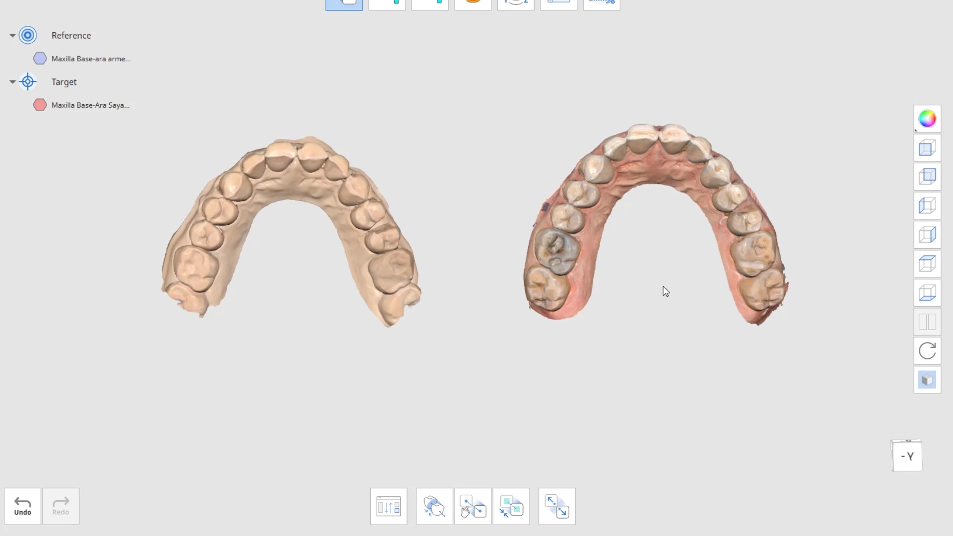
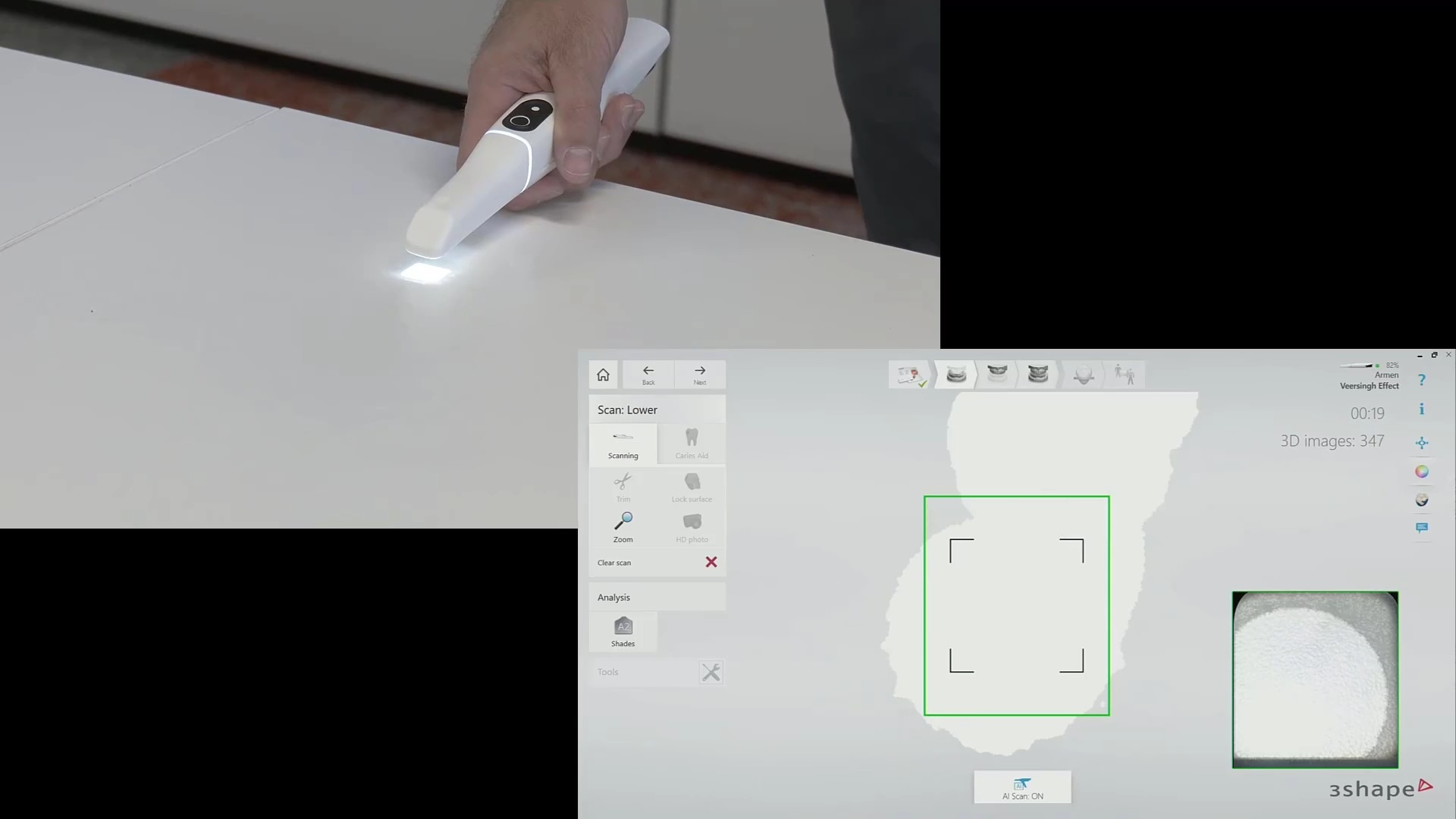
Additional Note: It’s essential to understand that in dentate cases, the properties of enamel can introduce errors. These errors arise from refraction, reflection, and light transmission through enamel. The asymmetric tooth morphologies can help your scanner stay on track. In edentulous cases, we face a different set of challenges related to the symmetric anatomy of the object being scanned, making it easy for the software to plot data points where they don’t belong.

Handheld vs. Desktop Scanners:

One of the video’s key takeaways is the difference between handheld and desktop dental scanners. Handheld scanners, like the ones we’ve featured, capture images and stitch them together to create digital models. Desktop scanners, on the other hand, take multiple views of a stationary object (also, with a wider field of view), resulting in higher accuracy and less room for errors. Understanding this distinction is vital in avoiding ‘Veersing’ and other scanning errors.

Examples of Scanning Errors:

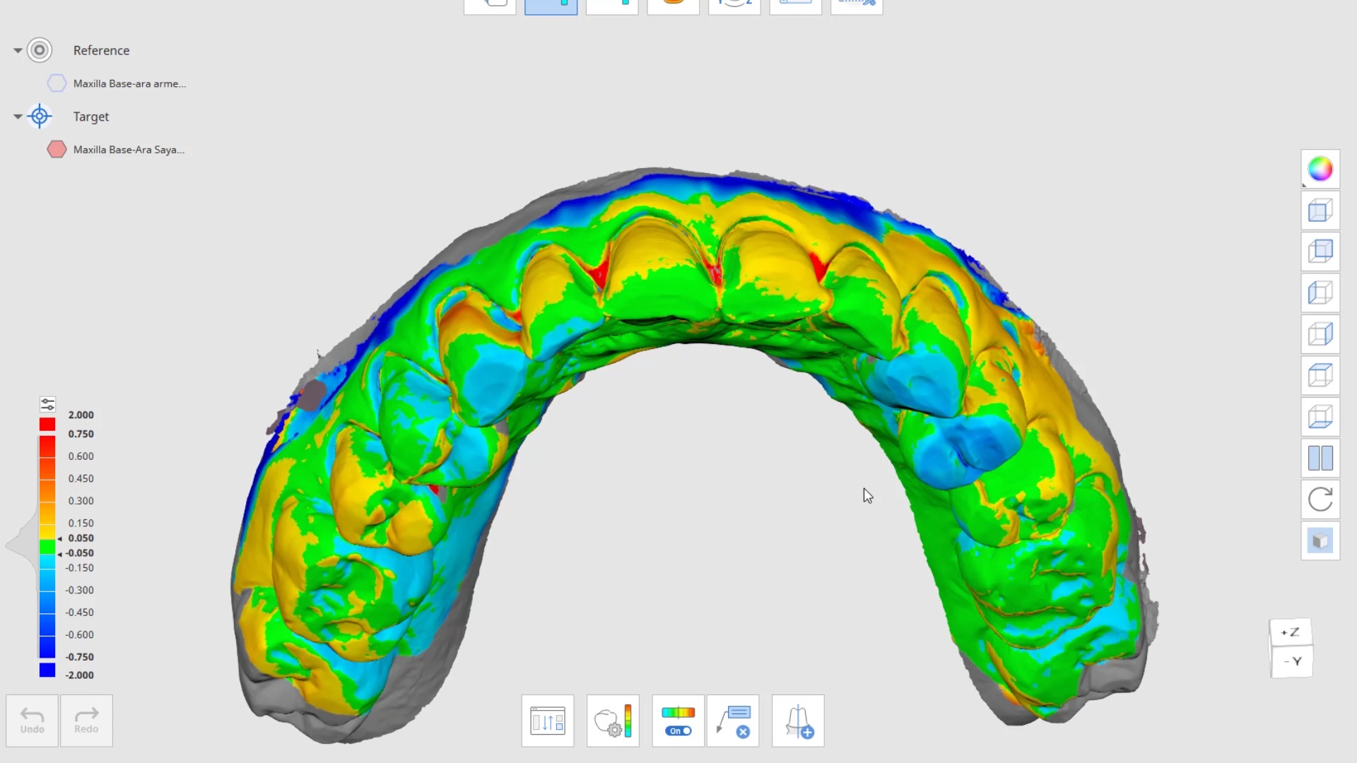
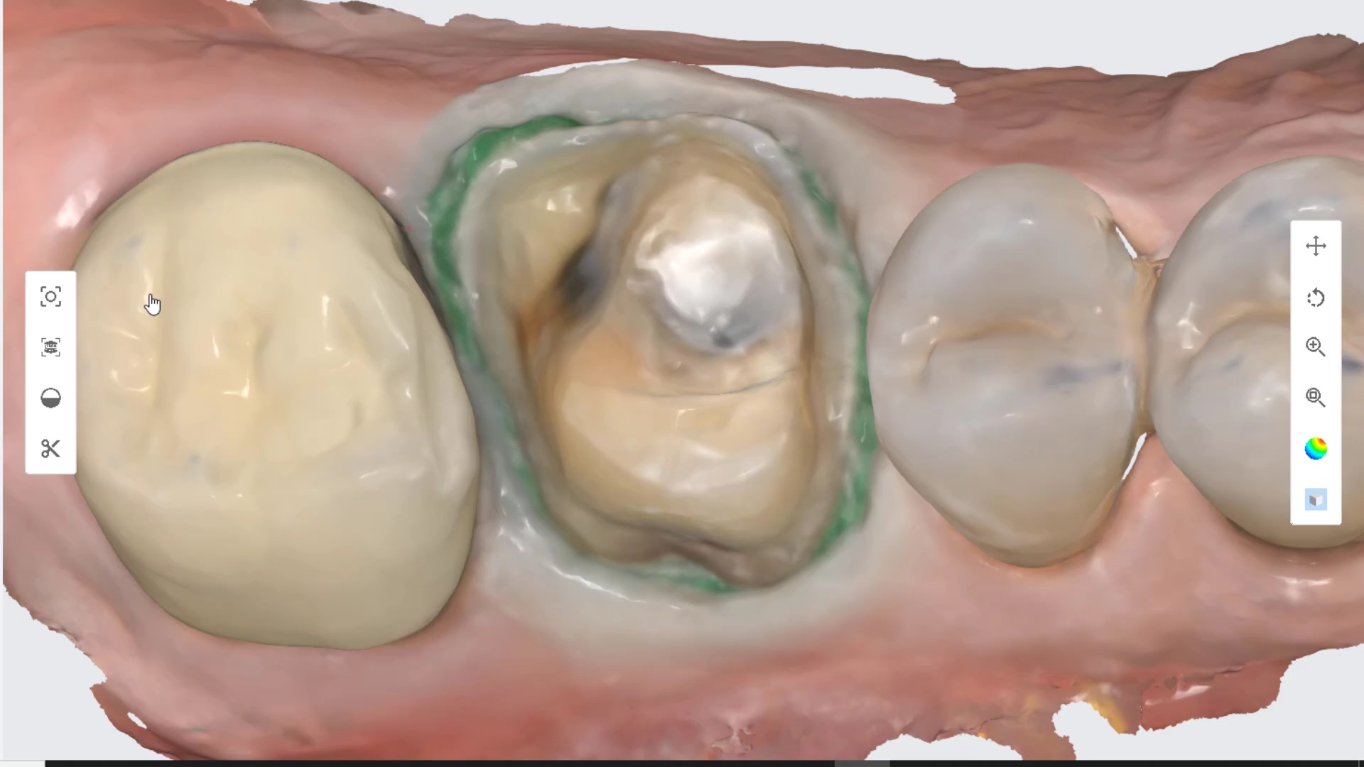
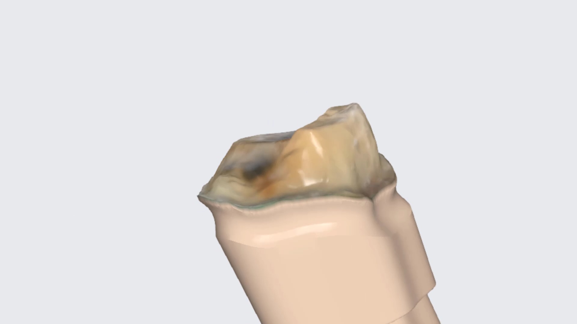
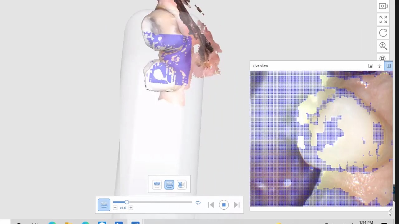
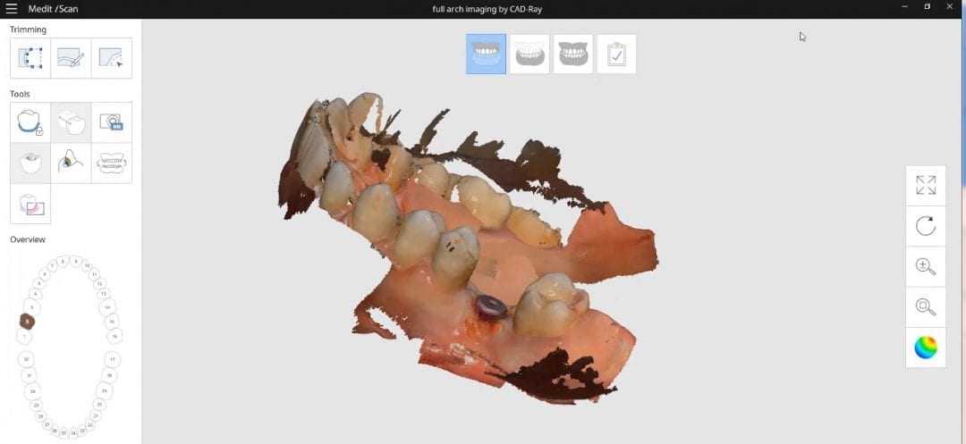
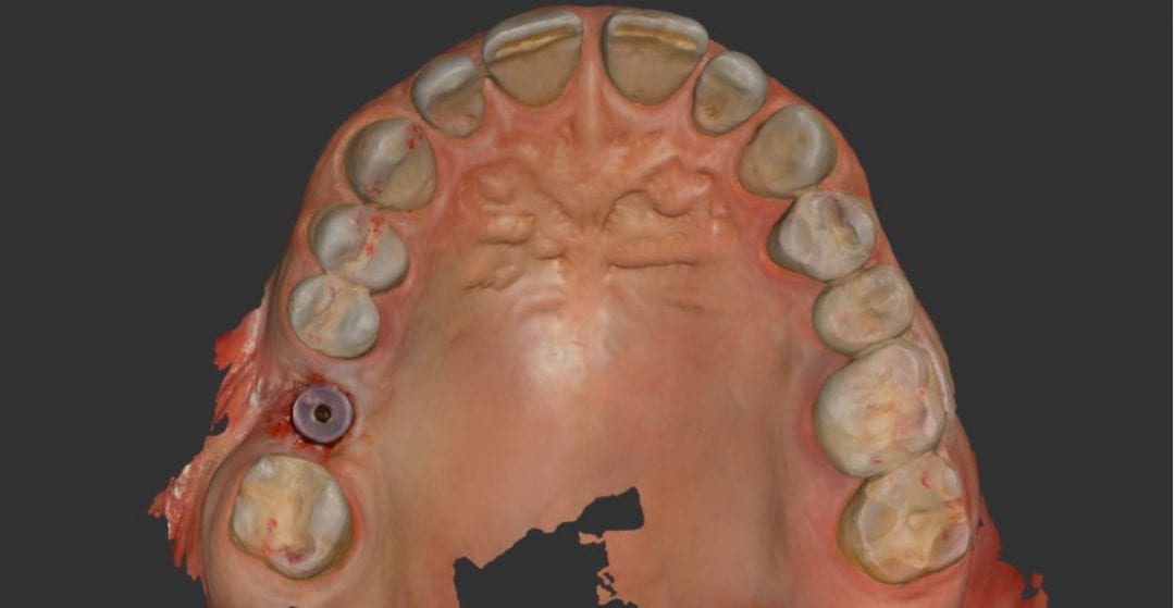
Dr. Mirzayan demonstrates the potential for scanning errors with real-world examples from the video. These examples provide a visual insight into how ‘Veersing’ can affect the accuracy of digital models created with these scanners.

Preventing Scanning Errors:

To prevent ‘Veersing’ and other scanning errors, proper scanner positioning is crucial. Maximizing the field of view is key, as it allows for more accurate data capture. Additionally, considering the virtues of desktop scanners, which inherently provide multiple views of stationary objects, is another important step toward achieving precision.

Importance of Accuracy in Digital Dentistry:

The overarching message of Dr. Mirzayan’s video is clear – accuracy in digital dental scanning is non-negotiable. Avoiding scanning errors is a cornerstone in treatment planning, leading to better patient outcomes and more precise dental work, and steps can be taken to reduce the potential for those errors.

Conclusion:

In a world where dental technology is constantly evolving, understanding and preventing scanning errors are paramount. Dr. Armen Mirzayan’s video is a valuable resource that highlights the nuances of ‘Veersing’ and scanning errors, providing insights for professionals to enhance their scanning processes. We invite you to watch the video and embark on a journey to a more accurate and precise dental practice.

Share your thoughts and experiences with us regarding digital dental scanning. If you have any questions or concerns about the scanners mentioned in the video or any other dental technology, don’t hesitate to reach out to CAD-Ray. For more information, please visit our website at www.cad-ray.com and watch Dr. Mirzayan’s video to gain a deeper understanding of ‘Veersing’ and the importance of avoiding scanning errors.

video
play-rounded-fill
The Veersing Effect
Posted on Leave a comment

Scanbodies and Edentulous Ridges

Scanning long span edentulous is a very likely culprit with rendering dimensionally accurate models because to the software and camera, the geometric shapes are too identical across the ridge.

The more we can disrupt the symmetry with large, short, non-reflective and asymmetric scanbodies, the more likely we are to keep the models accurate. This video presentation address some of those issues and how to overcome them.

video
play-rounded-fill
Posted on Leave a comment

Top Reasons Why Restorations from Digital Impressions Don’t Fit

Much like conventional impressions, digital impressions can render ill-fitting restorations.  It is imperative to figure out the source of the problem and to understand if it is a scanning or manufacturing issue.  We have compiled the top reasons for such errors in this article:

    1. If the restoration is manufactured on printed models, you should be alarmed! There are so many variables that can be introduced in additive manufacturing process that can lead to less than desired results.  Printed models should only be used after restorations are milled to assess contacts or to dial back bulky material to the margin on they die.
    2. Translucent / transparent enamel can lead to inaccurate scans.  This is most magnified when we work on conservative preparations made on enamel.  See this article to understand the troubles that can be introduced while scanning glass. aa

video
play-rounded-fill
Comparing Intra-Oral Scan with Printed Model

3. We highly recommend that the clinician places his or her margins as soon as they image the preparation. Oftentimes, labs only work with STL models instead of color models and this leaves a lot of room for open interpretation and errors.  Labs can then print the models to finish work like contacts and reducing margins they have bulked out to protect the material whilst milling.

video
play-rounded-fill
Mark your own margins
Posted on Leave a comment

Excessive External Light Source Can Alter Your Models

The overhead light source can distort your models and impressions. This particular doctor had excellent preparations and retraction but the light source kept flooding the dentition resulting in ill-fitting restorations. Our crew at CAD-Ray remotely logged in through teamviewer and was able to review the logged video while scanning and instantly noted the source of the error.

The Medit software and scanner have a setting that can warn you if there is too much excessive ligthing.

video
play-rounded-fill
Excessive light
Posted on Leave a comment

Fundamentals of Intra-Oral Scanners

Here are fundamental principles every dentist should understand about intra-oral scanners, how we can introduce errors, how to manage them, follow proper scan strategies, maintain accuracy across the arch and lots of other nuggets

Every ios hates moving parts (tongue and cheek)
Every ios hates dark areas like the oropharynx, black triangles, etc…
Every ios hates liquids (blood on the margins)
Every ios can “get off track” and form double images
Every ios struggles with highly reflective surfaces like emax, zirconia, and shinny metal.

The principles shown in the video address every one of these factors and how you can control them and get accurate arch scans in just a minute with the medit i500

I have tried virtually every ios out there and they all struggle with my mouth full of emax. the focal length of the medit from -1.5 mm to 17 mm is the largest variation on the market, and the way the medit handles highly reflective surfaces also outperforms every other ios.

In January, it will be in turbo boost mode when new software is released.

Posted on Leave a comment

Errors Introduced with Long Span Imaging and how to Correct Them

This case shows a full arch imaging where the model starts to go “off track”. To keep the explanation simple, the further away you get from multiple planes / heights of immobile structures, and the more you image in flat areas, you can inadvertently introduce errors in your models. You can see a sample case here in the video.

[videopress zqRzFmTj at=”8″ permalink=”false” hd=”true” autoplay=”true”]

There is a very simple solution, as you can see in this video. It entails consistently moving back to reliable landmarks to stitch new information to exisitng correctly. If you notice how we start imaging on the occlusal of the premolars and the molars, then we roll to the facial of the molars and the back around to the palatal of the molars. Then we sweep over soft tissue on the palate. But we don’t continue in that direction. We immediately return to the molar area so the software has landmarks it can recognize.

We then move the camera forward, image the palatal of the premolars, and then sweep back across the palatal midline. We repeat this back and forth movement to maintain a proper path for the software to recognize landmarks that do not move, in the equation. With this technique, you can scan a whole upper arch in just a couple of minutes and capture great details of the dentition and the soft tissue on the palate

[videopress VVEbeoWs permalink=”false” hd=”true”]

Here, you can see how the software rendered a perfectly accurate and detailed upper arch with the palatal vault captured without any errors.