I have been a customer of CAD-Ray for about a year now. We have purchased multiple items from Laura, including exocad software, a milling machine, and the Imetric4d. I can’t express enough how happy we are that we made those purchases through them. T...read moreI have been a customer of CAD-Ray for about a year now. We have purchased multiple items from Laura, including exocad software, a milling machine, and the Imetric4d. I can’t express enough how happy we are that we made those purchases through them. The customer service and the fast responses, (especially Laura) has been top notch! At the beginning, just like anything else in dentistry, we needed support and reassurance of the items acquired and all I have to say is a big thank you for an amazing customer service and support!!!! And did I mention that they offer really good financial options? Again, thank you! Looking forward for the next purchase. - The Inspired Dental team in Las Vegas:)read less - 8/31/2021
Jennifer Ebner
After using the medit i500 scanner at an office as an employee, I purchased it from Cad-ray,com specifically because of their good reviews and their large, concise training library of videos. I've used CEREC as an instructor at a dental school and bo...read moreAfter using the medit i500 scanner at an office as an employee, I purchased it from Cad-ray,com specifically because of their good reviews and their large, concise training library of videos. I've used CEREC as an instructor at a dental school and bought a used Planscan, Planmill for my private office. Both were hard to learn and the fees and cost associated with training were a bitter pill to swallow after the costs to purchase. The ease of seating restoration was instant. The scanning learning curve is quick. I have easily added occlusal guard scanning, and implant scanning and will be trying a digital denture soon.
The purchase was easy, the product came quickly and the set-up videos guided me through very necessary adjustments to my Laptop I never knew I needed. Learning the new features with each FREE update is a treat. The DIY option was an extra bonus as the $2000 rebate came without having to ask for it. Cad-ray has delivered more than I expected with the purchase of the fantastic Mediti500 scanner.
I've included my cart set up. purchased from Amazon for $68. Its stable, has room for the necessities. I've taken it home and back to the office half a dozen times and it transports the scanner safely. It also keeps the cords from being run over. I haven't taken the included 2 day course but look forward too it after Social Distancing mandates are lifted.read less - 4/25/2020
I have been very happy with the Medit I purchased from Cad Ray. Everyone has been very helpful and quick to respond to my questions. They have an awesome video library for the medit, I am new to scanning and have really enjoyed the tutorial videos. - 6/16/2020
John Eum
Love all the staff there. Great support and instruction from Armen, Laura, Damien. We are very grateful for Kaila who has been incredible in getting us going - very friendly, professional and responsive. Thank you! - 7/03/2024
Michael Martinsen
Honestly the best service that I've received in dental tech support. I had originally purchased a scanner with another supplier and was underwhelmed with the support I received. I switched over to CAD-Ray for my second scanner and have never been let...read moreHonestly the best service that I've received in dental tech support. I had originally purchased a scanner with another supplier and was underwhelmed with the support I received. I switched over to CAD-Ray for my second scanner and have never been let down. They are prompt and exceptionally knowledgeable about the products they support. Thank you CAD-Ray for keeping my practice moving smoothly!read less - 8/27/2021
Meridien Dental
Got my Medit scanner from here. Purchase went very smoothly. Best part is the on going support. Cad-ray team is great with training and ongoing help if your scanner has hiccups here and there with various updates. - 12/11/2023
Mark Gerome
When I bought my first scanner a few years ago I was blown away by CAD-RAY’s support! They do a great job of getting you and your team ready to go with it. Frank D. has been a huge help with getting us confident with our scanners and making sure we a...read moreWhen I bought my first scanner a few years ago I was blown away by CAD-RAY’s support! They do a great job of getting you and your team ready to go with it. Frank D. has been a huge help with getting us confident with our scanners and making sure we are taken care of. He and his colleagues at CAD-RAY are always a responsive to our calls. Thank you!read less - 2/21/2023
Armen Mirzayan
Are you happy with the support and training you have received with our team at CAD-Ray.com?
Our business grows much like yours, with referrals and recommendations. We would greatly appreciate a quick review / testimonial
NOTE: ...read moreAre you happy with the support and training you have received with our team at CAD-Ray.com?
Our business grows much like yours, with referrals and recommendations. We would greatly appreciate a quick review / testimonial
NOTE: All testimonials are submitted by verified owners.read less - 5/08/2021
I have had excellent , prompt , customer service and support from the entire team . Especially from Frank DeLuca as I move along the learning curve. His patients and expertise is much appreciated - 1/24/2023
Kurt Adamson
I purchased My first Medit i500 in Sept 2019, fairly easy to set up and learn. I was able to follow the instructions and videos without a problem. I learned tips and trick along the way by following the facebook groups. Anytime help was needed the g...read moreI purchased My first Medit i500 in Sept 2019, fairly easy to set up and learn. I was able to follow the instructions and videos without a problem. I learned tips and trick along the way by following the facebook groups. Anytime help was needed the group at Cad-Ray were quick and sincere finding a solution to problems. I ended up purchasing 2 more i500's in Dec 2019 for my other 2 offices. I didn't think I would enjoy the digital workflow as much as I do. If you on the fence like i was, just do it. get on the facebook groups and post the tips you learn along the way.read less - 1/17/2020
We had an outstanding experience purchasing two Medit scanners. The delivery was timely . The onboarding clean. Laura was dazzling with her acumen and quick solutions . We highly recommend the investment in Cad Ray .Pobanz Orthodontics , Ogden Utah :...read moreWe had an outstanding experience purchasing two Medit scanners. The delivery was timely . The onboarding clean. Laura was dazzling with her acumen and quick solutions . We highly recommend the investment in Cad Ray .Pobanz Orthodontics , Ogden Utah :)read less - 12/10/2021
Jared Gustafson
One of the best companies to work with for dental tech needs. They have a vast video training library that makes the transition easy to digital dentistry. I’ve recommended a few of my colleagues purchase the medit through them. It’s been a great year...read moreOne of the best companies to work with for dental tech needs. They have a vast video training library that makes the transition easy to digital dentistry. I’ve recommended a few of my colleagues purchase the medit through them. It’s been a great year!read less - 6/16/2020
Peipei Yu
I purchased a Medit scanner in August and I am very happy with the scanner. I was trained on an Omnicam in dental school and used Omnicam for 4 years in my associateship. The speed and quality of the Medit scanner is comparable to a CEREC scanner but...read moreI purchased a Medit scanner in August and I am very happy with the scanner. I was trained on an Omnicam in dental school and used Omnicam for 4 years in my associateship. The speed and quality of the Medit scanner is comparable to a CEREC scanner but you can’t beat the price of the Medit! I would highly recommend this if you are a beginner or pro in digital dentistry!read less - 9/21/2020
Kyle Coffin
Bought a medit 6 months ago and customer service has been nothing short of amazing. Always very responsive and helpful. Great scanner, too! - 6/16/2020
Hieu Pham
Been super happy with service from this company. Very responsive, really passionate about their work. - 7/16/2020
Blake Ferando
I purchased my medit in April of 2019 from Cad-Ray. The support offered is second to none, and the training videos are some of the best out there. Add to that a great support team that is fast to answer questions and issues, its hard to beat Cad-Ray. - 6/19/2020
Mark Gaches
Excellent company to work with. Best customer service and technology available. Can't recommend enough! - 5/05/2021
Emerson Gower
Nothing but great things to say about Cad-Ray! Excellent customer support from Laura after the purchase of our Medit i500 and the cart from Damien. I’ve been very impressed with the Medit in restoring crown and bridge, as well as fixed hybrids, and...read moreNothing but great things to say about Cad-Ray! Excellent customer support from Laura after the purchase of our Medit i500 and the cart from Damien. I’ve been very impressed with the Medit in restoring crown and bridge, as well as fixed hybrids, and learning more of its capabilities each day. We have intentions to buy a second Medit shortly and will definitely be using Cad-ray again.read less - 12/13/2021
Nisarg Parikh
Absolutely a phenomenal company. Everyone you engage with is extremely knowledgeable and helpful.I’m sure they get the same questions day in, day out but they are happy to help you without being condescending. The entire team has been helpful through...read moreAbsolutely a phenomenal company. Everyone you engage with is extremely knowledgeable and helpful.I’m sure they get the same questions day in, day out but they are happy to help you without being condescending. The entire team has been helpful through phone calls, the Cad-Ray FB group, and recorded webinars. Jonathan helped me get a great deal, Laura was awesome on the webinar, Damien is always answering questions on FB, and Armen had a great in-person presentation in Dallas. 100% recommend.read less - 4/22/2022
Ernesto Carmona
When I decided to purchase a digital scanner I decided on user experience. They have provided the best experience I could have asked for, from online support, Facebook support, and in person courses. There is no better team to buy Medit from period...read moreWhen I decided to purchase a digital scanner I decided on user experience. They have provided the best experience I could have asked for, from online support, Facebook support, and in person courses. There is no better team to buy Medit from period. Do yourself a favor and buy from them if you are in the market for an intraoral scanner.read less - 6/16/2020
Otto Herod
I received my Medit scanner a couple months ago from Cad-ray, and I can't express enough how awesome it is. I have done a ton of research and used the latest IOS from one of the big guys, and due to many software issues I was able to return that mach...read moreI received my Medit scanner a couple months ago from Cad-ray, and I can't express enough how awesome it is. I have done a ton of research and used the latest IOS from one of the big guys, and due to many software issues I was able to return that machine. And thank God, because it was so over priced and came with a $300/month support fee forever! The Medit scanner is as good or better than that one when it was working properly, and for the price it's a no brainier.read less - 9/17/2020
Mehryar Ebrahimi
Great costumer service. I needed a part for my i700 and they were able to ship overnight. No down time. - 5/06/2022
Jason Ehtessabian
Fantastic! Scanner went down after a few years, and they had a new one in my office the next morning. Highly recommended. - 8/26/2021
ycsongdds
I have had zoom training with Andy today. I can honestly say that he was the best. He was patient and very throughal. I learned a lot I would ask him again when I need support again Thanks Rodger - 3/15/2023
Barton Davis
Every time I need support for my scanner, the Cad-Ray team is there to help. I recently had a question on how to manipulate a scan and export it back to Medit Scan. Damien logged in and helped me out. Problem solved in under five minutes. Awesome ser...read moreEvery time I need support for my scanner, the Cad-Ray team is there to help. I recently had a question on how to manipulate a scan and export it back to Medit Scan. Damien logged in and helped me out. Problem solved in under five minutes. Awesome service. Thanks Cad-Ray support team!read less - 3/04/2022
Lake Shore Dental of Tempe
Absolutely the best customer service I have ever seen. I couldn't be happier with the service received. I won't be going anywhere else!! Thank you again. - 2/01/2022
Erica Zolnierczyk
Purchased the Medit right before the COVID shut down, which gave us time to train on it by watching all the awesome videos CAD-Ray provides. If I couldn’t figure something out, our questions were answered quickly. Now we’re back and using it like cra...read morePurchased the Medit right before the COVID shut down, which gave us time to train on it by watching all the awesome videos CAD-Ray provides. If I couldn’t figure something out, our questions were answered quickly. Now we’re back and using it like crazy. It’s quick, my assistant picked it up quickly and my cases have been coming back perfect. Super happy with the Medit!read less - 6/16/2020
Christopher Chin
This was my intro into intraoral scanning and I did research for quite awhile. The i500 and the team at Cad Ray are top notch. The company has done such a great job putting this scanner in the ranks of the premier ones. With constant updates it gets...read moreThis was my intro into intraoral scanning and I did research for quite awhile. The i500 and the team at Cad Ray are top notch. The company has done such a great job putting this scanner in the ranks of the premier ones. With constant updates it gets better and better (though I’m still behind on updates). My crowns have never been better. They drop right in with no models. I didn’t believe it but I’m loving digital dentistry. I can’t wait to go to the class after COVID calms down!read less - 6/16/2020
It's so nice to enter the digital age for scanning! I've been taking impressions since 1975. Medit i700 is amazing and our #1 supporter, Laura has been with us every step of the way. I truly don't know what I would do without Laura and Nick in tec...read moreIt's so nice to enter the digital age for scanning! I've been taking impressions since 1975. Medit i700 is amazing and our #1 supporter, Laura has been with us every step of the way. I truly don't know what I would do without Laura and Nick in tech support. They definitely need a raise! Yes, we had a couple of glitches (mostly operator error) but they were there with us all the way. The detail that the scan gives, whether it's a crown, bridge, full mouth scan for patient review and now many scans for NTI's is truly amazing! We recommend it highly to all who are interested! I am proof that as a dental assistant who's 64, you CAN teach an old dog new tricks!!!read less - 12/09/2021
Chad Gardner, DDS
Can not find a single negative thing to say about my transactions with CAD-Ray. Great people, absolutely unbelievable customer service, and the best products.They stand behind the products they sell as strongly as the manufacturers do. - 4/27/2022
Luciana Bretz-Pavie
Wayne Glassoff is the go-to guy here! The BEST!!! He is always on it, he helps figuring out exactly what you need, the best way possible. And fast! Great working with CAD-Ray, they have pretty much everything when it comes to digital dentistry. And t...read moreWayne Glassoff is the go-to guy here! The BEST!!! He is always on it, he helps figuring out exactly what you need, the best way possible. And fast! Great working with CAD-Ray, they have pretty much everything when it comes to digital dentistry. And they give you a damn good support too. Jessica Knott is awesome… training and support, just the BEST! Very satisfied customer here!read less - 2/05/2023
Ma. Teresa Santana
Best support and customer service ever! My scanner is long past warranty and they still answer all my questions. When it's time to upgrade I'll be buying from them again. A friend bought same scanner from another vendor and got zero support. I had to...read moreBest support and customer service ever! My scanner is long past warranty and they still answer all my questions. When it's time to upgrade I'll be buying from them again. A friend bought same scanner from another vendor and got zero support. I had to help them. Told her next time buy from Cad Rayread less - 5/10/2024
Norman Knowles
I have had the original iTero, a Trios 3, and a Carestream CS3600. A staff member broke the lens on the Trios and while waiting 3 weeks for an RMA to send it to Poland for six weeks to get it repaired, Carestream was sniffing around and suggested tha...read moreI have had the original iTero, a Trios 3, and a Carestream CS3600. A staff member broke the lens on the Trios and while waiting 3 weeks for an RMA to send it to Poland for six weeks to get it repaired, Carestream was sniffing around and suggested that I trade in my Trios so I did which began a 4 month nightmare with their piece of junk scanner. Absolutely awful customer service from both 3Shape and Carestream. I needed a scanner and had heard great things about it the Medit i500 at the Florida Academy of Coesmetic Dentistry so I got one. It works really well and Medit keeps adding new and useful features and their architecture is completely open. Two weeks ago, my i500 died so I contacted Cad-Ray surrport and I had a new unit in my office 24 hours later! That's absolutely unheard of in the industry!!! Both Cad-Ray and Medit have positioned themselves to be industry giant killers and they're doing it. The i500 is a phenomenal scanner at a phenomenal price point and service and guidance at Cad-Ray is just plain excellent.If you are thinking about getting a scanner, do it! Just contact Cad-Ray and go for it. You will only be happy about your purchase! Highly recommended!read less - 3/09/2021
Deryck Pham
best customer service on the planet. very easy to use. and always fine tuning to make the user experience better. - 6/16/2020
Alderman Dental
Frank DeLuca from CAD-Ray has been absolutely awesome from day one. Readily available and very knowledgeable to provide support when needed. - 1/25/2023
H C
I got my 3Dshape scanner, great support , my training with Destany was excellent. Great team!! - 2/15/2024
Suzanne Stock
Excellent experience, customer service has been stupendous! - 11/20/2023
W Timothy Brooks
Excellent fast color scanner. A great improvement over my old proprietary scanner. Easy to learn software and great free technical support. - 6/18/2020
Armen created series of these very helpful videos. They helped us tremendously in breaking into the new scanner we purchased. No matter you just got into digital dentistry or you are quite experienced with different fields of data acquiring or treatm...read moreArmen created series of these very helpful videos. They helped us tremendously in breaking into the new scanner we purchased. No matter you just got into digital dentistry or you are quite experienced with different fields of data acquiring or treatment planning, you will find those being tremendous help.read less - 5/24/2020
We purchased Medit i-500 with CAD ray last December and customer support is great.One of best thing for our practice is having intra oral scanner ,works great . Laura was very patient with our learning curve. - 12/10/2021
Page Barden
I have just recently purchased the Medit i700, and although it is "on its way" I have to comment on the support that I have already received, especially from Nick. He walked me through the benefits and features of the scanner and the transaction was...read moreI have just recently purchased the Medit i700, and although it is "on its way" I have to comment on the support that I have already received, especially from Nick. He walked me through the benefits and features of the scanner and the transaction was extremely easy. I can only think that the rest of the onboarding will be the same and in six months I will revisit this site and be extremely grateful I made the purchase.read less - 12/09/2021
fantastic support, I always ask random, specific to me, could be found on a training video, questions and they always quickly tell me how to fix my problem. - 6/16/2020
Lance Timmerman
Implant Planning Services Review I Am VERY experienced with "another system" but thought I would give CAD-Ray a try. At at LEAST 1/3 the fee, why not?
VERY happy. Fit GREAT, surgery was uneventful (the best kind) and I am a believer! This is a great company and great PEOPLE to...read moreI Am VERY experienced with "another system" but thought I would give CAD-Ray a try. At at LEAST 1/3 the fee, why not?
VERY happy. Fit GREAT, surgery was uneventful (the best kind) and I am a believer! This is a great company and great PEOPLE too! 5 stars aren't enough. Just like the Burj Al Arab, this is 7 star!read less - 3/03/2015
Samuel Schmidt
I purchased a SprintRay through them. I'm new to the 3D printer realm and they've been great with answering my questions and getting me set up. I've just really have been impressed with the quick responses, its much appreciated, thank you! - 4/20/2022
Michael White
Everyone I have dealt with during the process of buying my new N4 VHF mill (which by the way is fantastic) last year to buying a new iMedit 500 scanner. Having the open type system has been a God send. No more held hostage by the 2 main systems us ...read moreEveryone I have dealt with during the process of buying my new N4 VHF mill (which by the way is fantastic) last year to buying a new iMedit 500 scanner. Having the open type system has been a God send. No more held hostage by the 2 main systems us same day crown dentists have had to deal with for years. Cad-ray made the financing a snap and the post customer support is unbelievable. Thank you everyone at Cad-Rayread less - 5/12/2020
Cristy Duval
Kaila is an incredible Rep! Hope Cad-Ray knows what a great employee they have. Thank you Kaila! - 12/16/2021
Scot Bruce
We found the digital training very informative - 8/20/2021
Roopam Garg
I purchased Medit scanner few weeks ago . Frank and Nick have been really excellent answering my questions promptly. The scanner itself is wonderful with no back fees , customer service is top notch. The entire team works fast and helps you out. I st...read moreI purchased Medit scanner few weeks ago . Frank and Nick have been really excellent answering my questions promptly. The scanner itself is wonderful with no back fees , customer service is top notch. The entire team works fast and helps you out. I strongly recommend this scanner!read less - 6/17/2020
These guys are ALWAYS there to help if you get stuck. I’ve been happy with the mill (coritec one) and scanner (i500) I bought from them 2 years ago. I’d do business with them again. - 7/01/2021
Dennis Wong
Best support ever. They are super responsive when a problem occurred, and immediately set up a support ticket with the vendor who contacted me within the hour to help me to solve the problem. - 7/27/2023
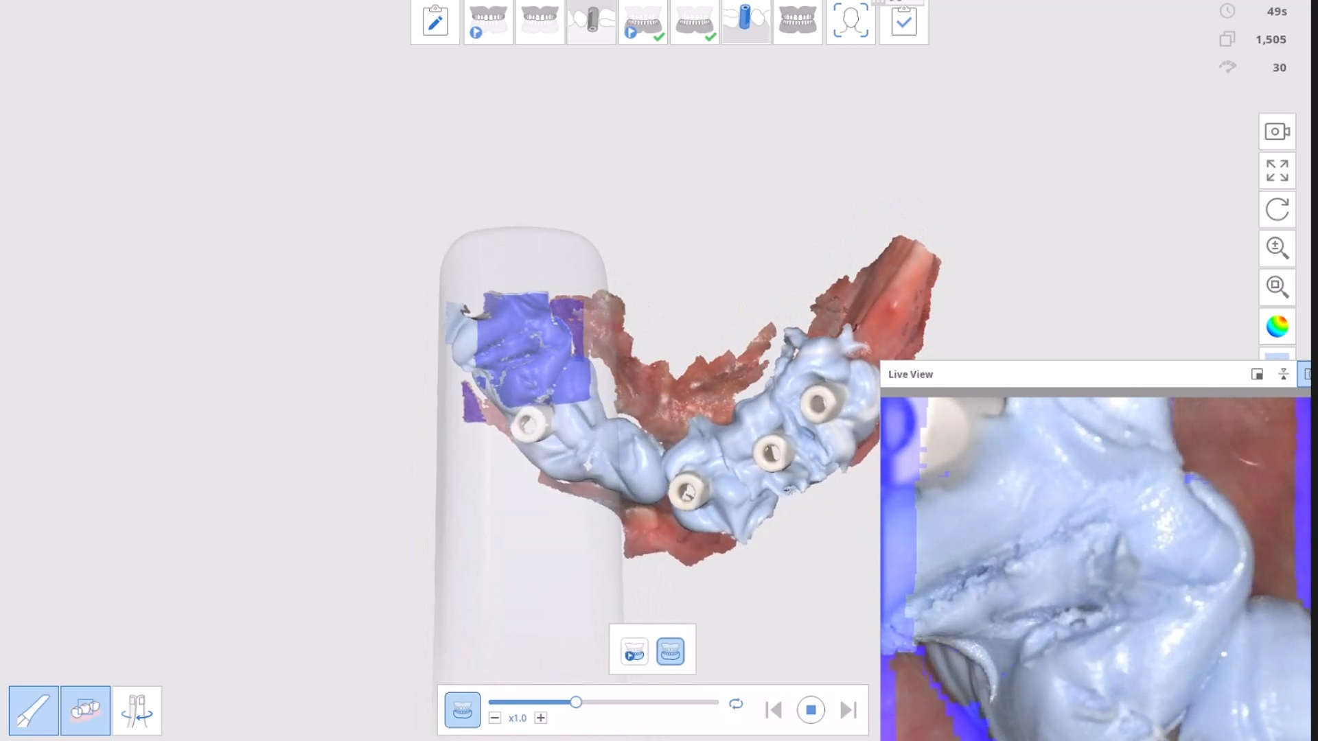
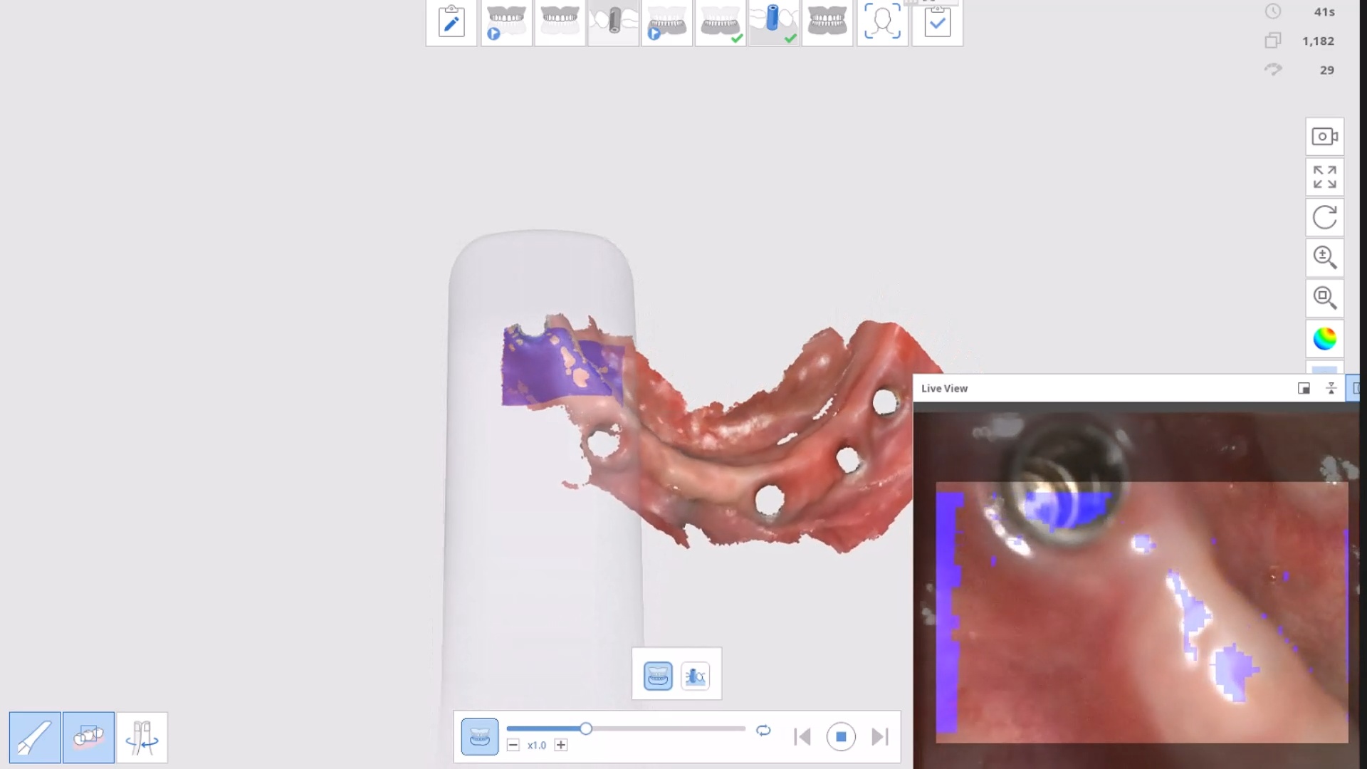
Scanning long span edentulous is a very likely culprit with rendering dimensionally accurate models because to the software and camera, the geometric shapes are too identical across the ridge.
The more we can disrupt the symmetry with large, short, non-reflective and asymmetric scanbodies, the more likely we are to keep the models accurate. This video presentation address some of those issues and how to overcome them.
here is a list of why the Medit Artificial Intelligent Implant Suprastructure Identification System is significantly more advantageous over all other cadcam systems.
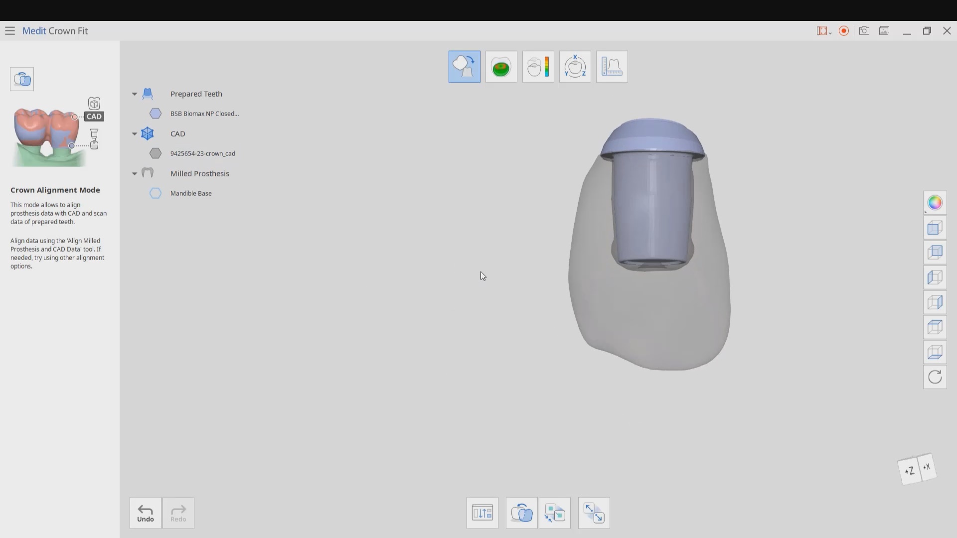
It s technically a crown and bridge case and the implant location or timing does not matter
You can find margins outside the mouth! See the first video to appreciate the significance of this
You don’t have to deal with retraction or hemostatis at all
You don’t have to worry about sprue position. Many other systems force the placement of the sprue to a specific location often making the case more difficult to manage than necessary
you are not limited to just a few implant lines
you don’t have to worry about location of anti rotational notch
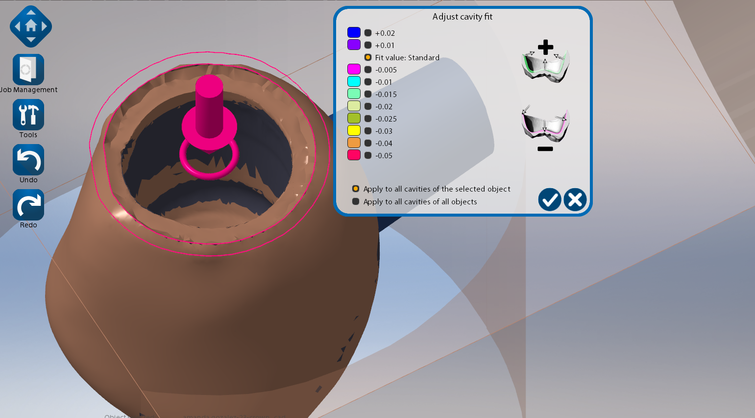
you can digitally alter the prep and get a virtual reduction coping in cad
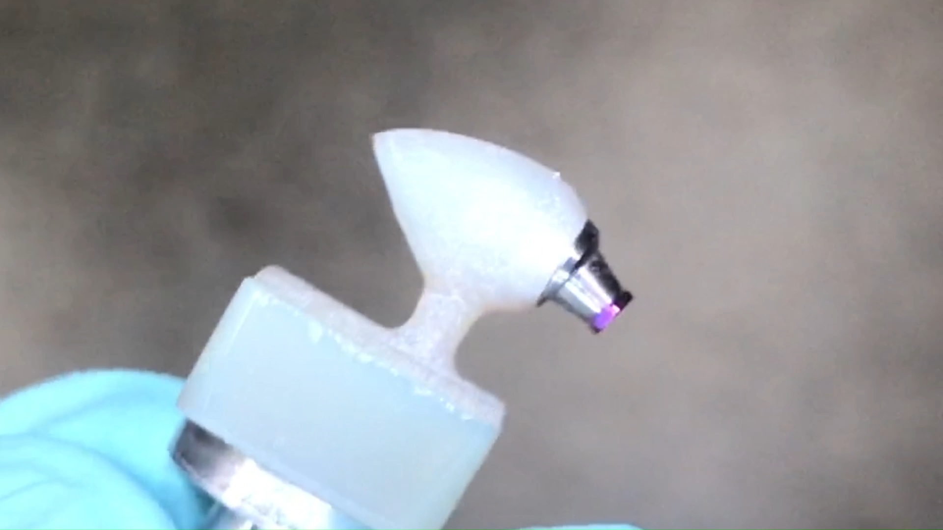
Use any restorative block you want. There is no need to order special blocks with pre-fabricated access channels and keep a large inventory of many colors. Your regular block inventory will suffice. Just make sure the top of the tibase is wider than the diameter of the drill used to mill out the intaglio. Also, the CAM and the milling machine determine the exact product and different settings maybe utilized to give you relief off the walls. Some will even remove the antirotational notch because the adaptation is so tight, the restoration will not rotate due to the tall walls of the tibase
You can check the fit outside the mouth on the same tibase or a one you keep chairside for every case to let you know that if you are not seating, it is clearly a contact or contour issue as opposed to an intaglio issue.
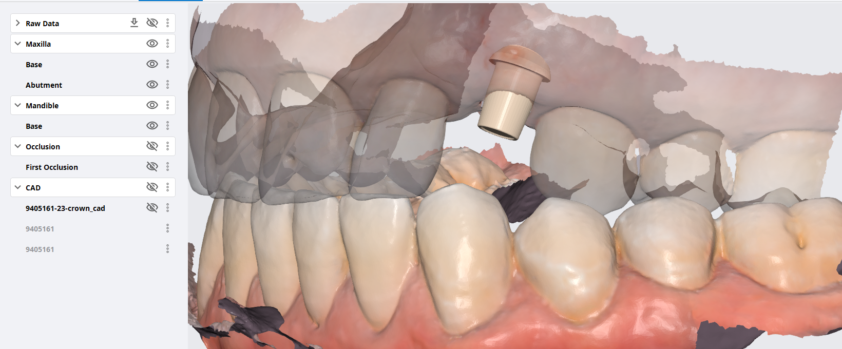
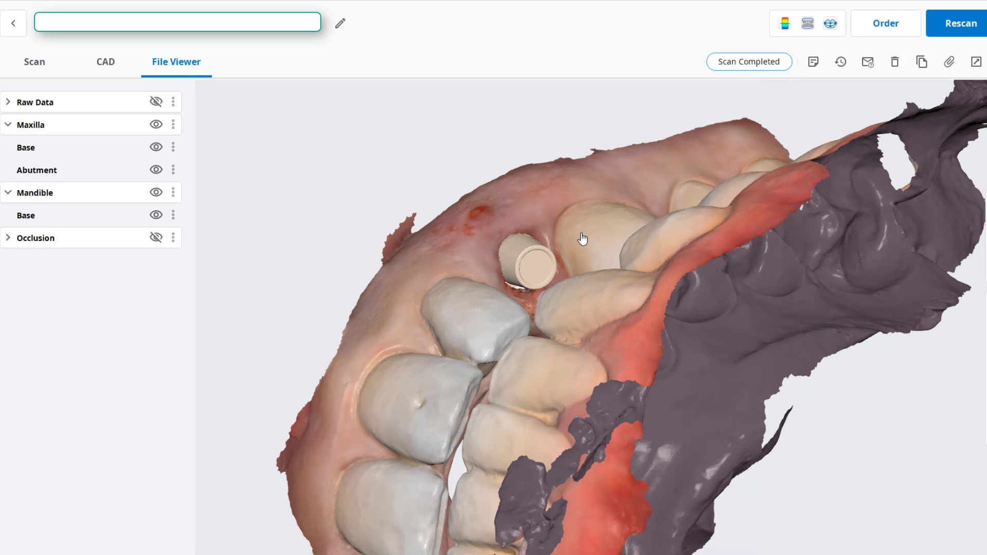
A cool new feature in the upcoming Medit i500 is the opportunity to capture deep areas that are out of the camera’s focal length (-1.5 to 17mm). This usually happens in complex implant cases, or in this case where the anterior four teeth were traumatized.
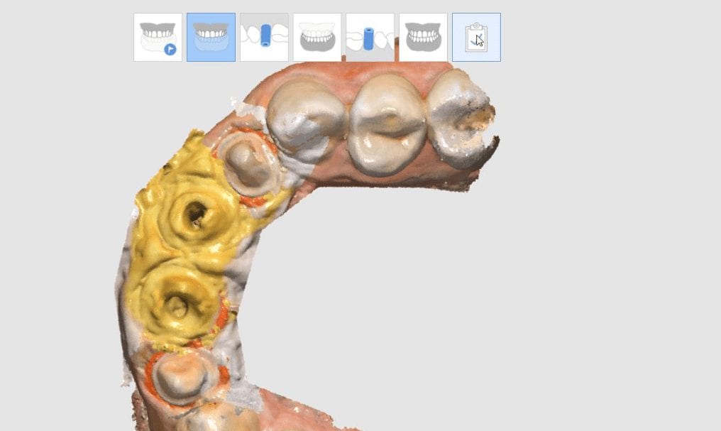
With very little tooth structure remaining, a custom post and core was required to restored the dentition with a very guarded prognosis. After root canal therapy, the chamber was accessed and a conventional impression was taken.
Separate from this, a clinical digital impression was taken of the temporaries, the opposing and the buccal bite. The margins of the preps were protected and the chamber was deleted / cropped. As you can see, there are hollow areas in the depth of the chambers where the topography was outside the focal length. Scanning the impression as a negative gives easy access to the depths of the chamber, allowing you to form a model that is well outside the imagine range of the neighboring teeth.
The software allows you to image intra-orally and then allows you to fill in the voids by imaging the impression instead.