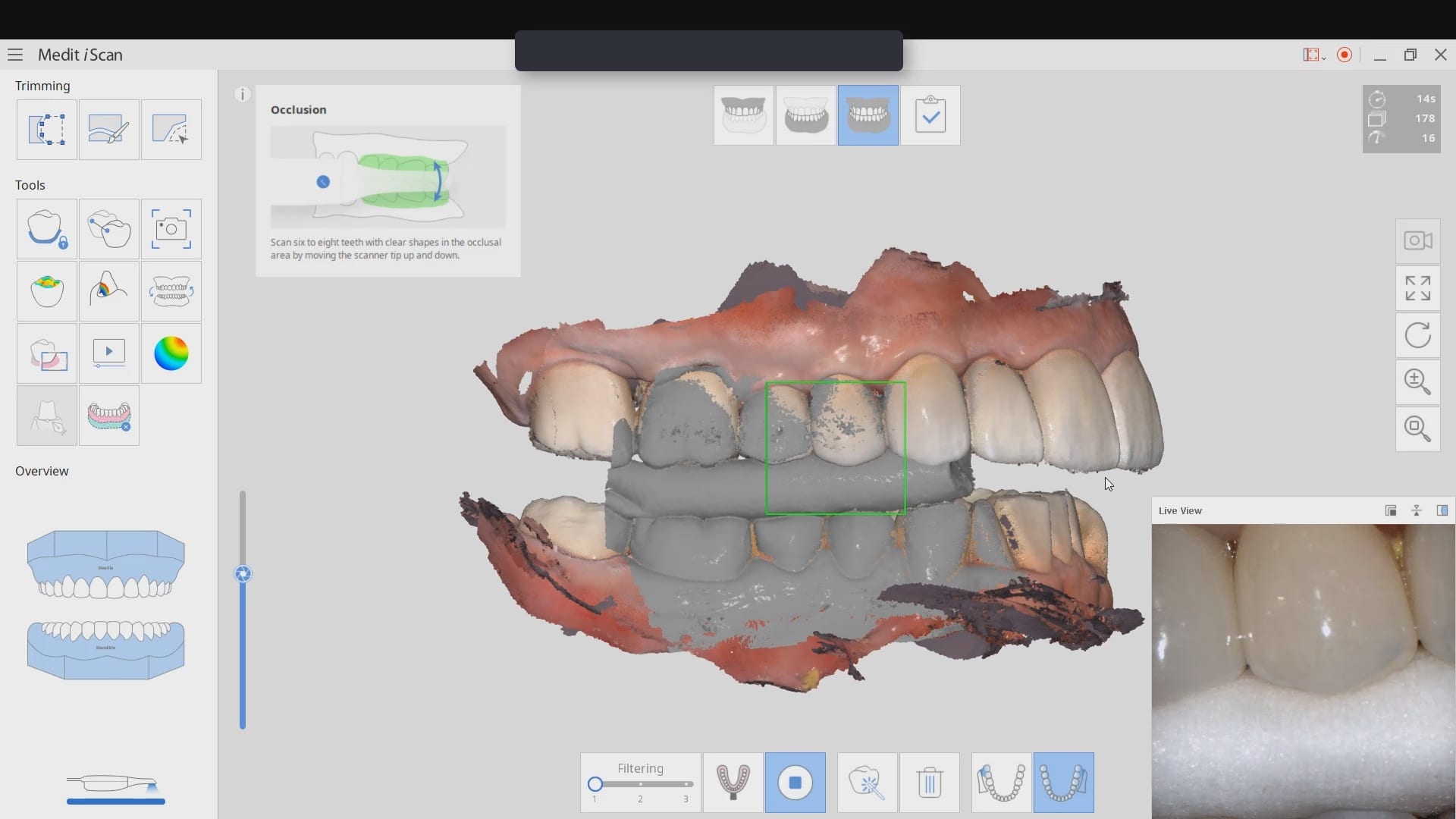


Many intra-oral scanners struggle with “black triangles” where there is tissue recession. Another area that is difficult to capture is the distal of second molars. Here is a video that shows a live scan with the Medit i500 that starts from the mid-line and images all the way to the second molar, captures the distal of the second molar, crosses over to the palatal and scans from to the contra-lateral second molar.
Once the distal of the second molar is captured the camera is rolled to the buccal, and the imaging continues to the midline, on the facial. The second video shows the upper and lower arches articulated together
[videopress GpFCcORn hd=”true” loop=”true” autoplay=”true”]