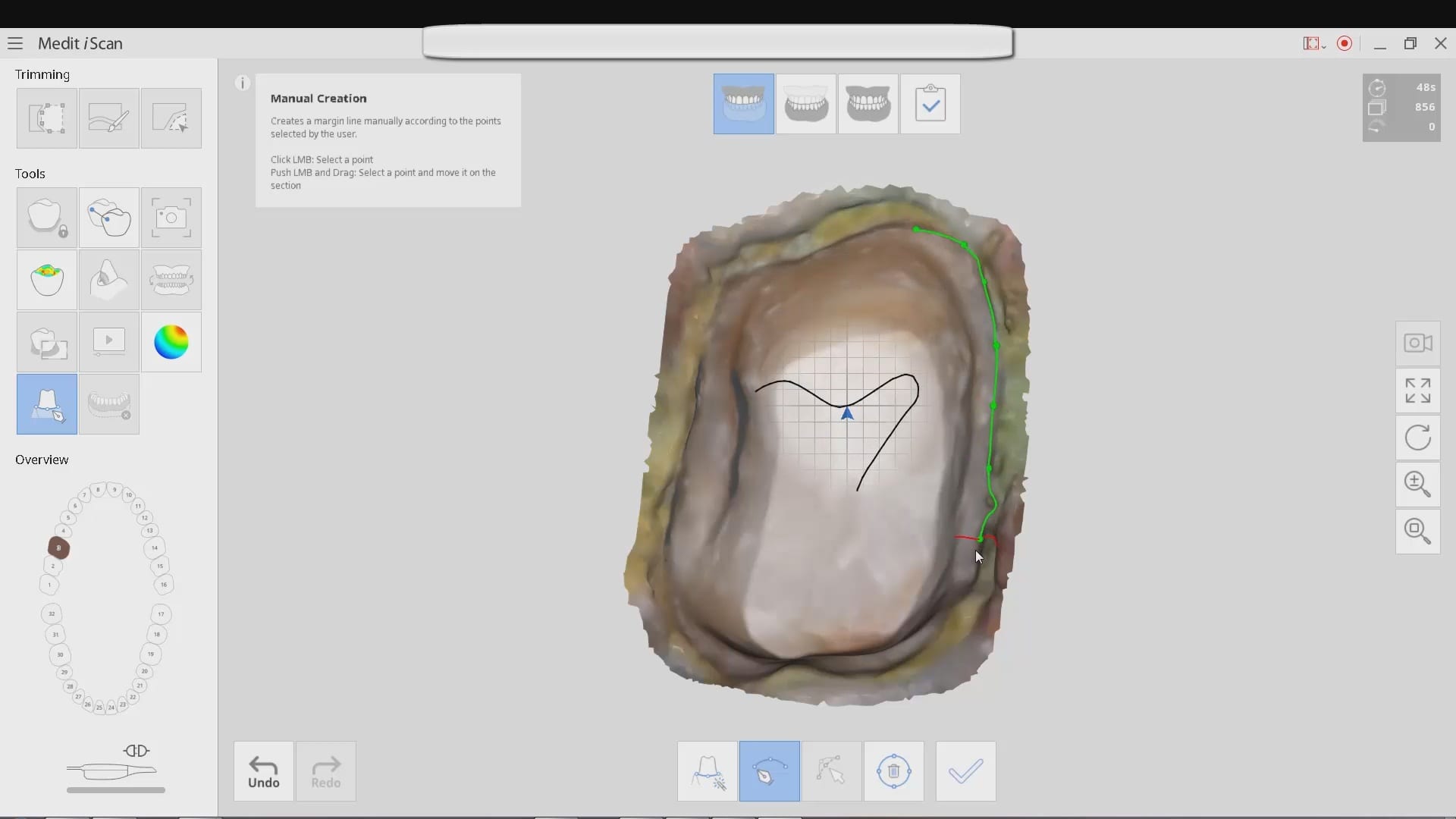


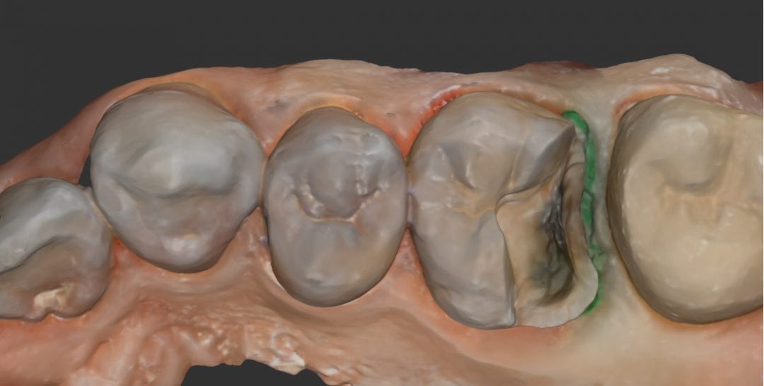
This is a case presentation of a onlay preparation on an upper first molar. The area was isolated with the optragate and the isolite after anesthesia. After a little refinement, a green colored retraction cord was placed to displace the tissue from the gingival margin.
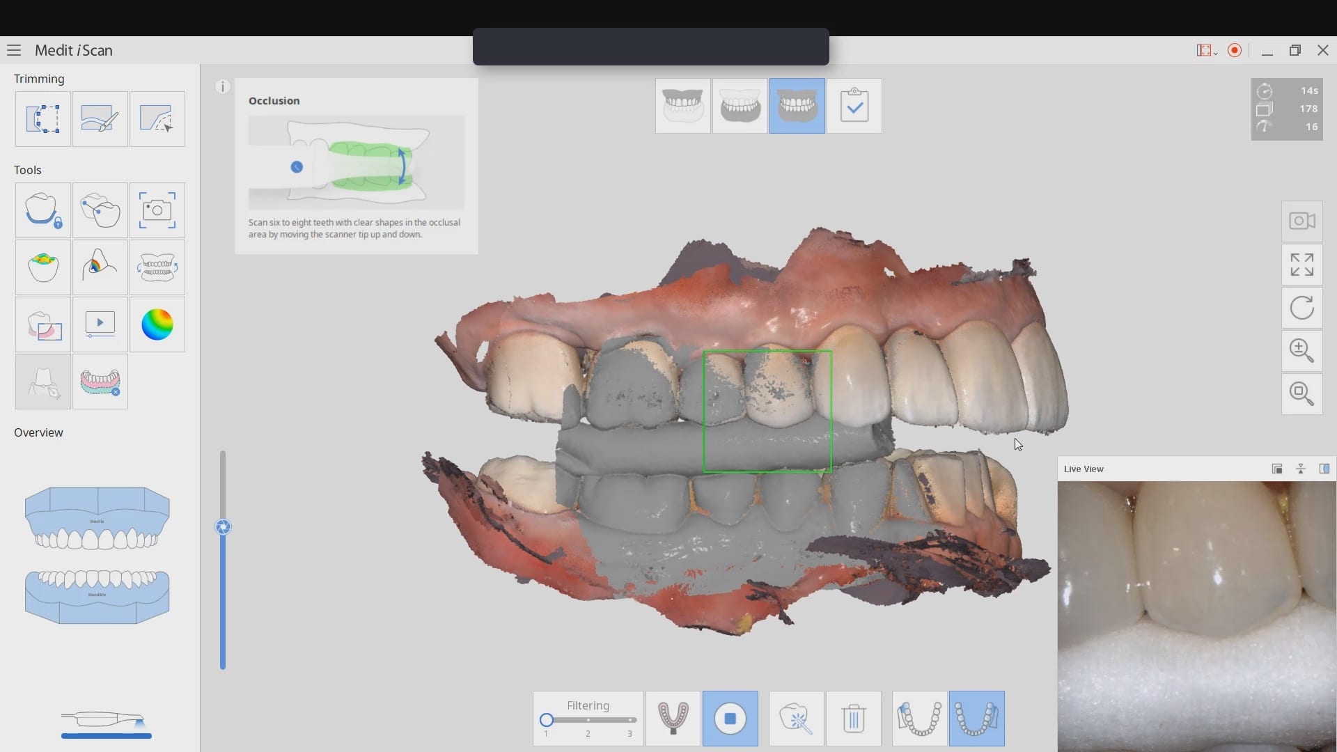
The attached video is unedited to show you exactly how long it takes to image a quadrant with the Medit i500 intra-oral scanner. You see an image counter and a clock in the upper left corner. First the prepared arch was captured. Notice how the camera is angled to capture all of the contact area on the second molar, below the height of contour. There are some areas that are not clinically relevant and less time was spent imaging those landmarks (palatal and lingual areas that will not have an impact on the restoration design).
After the upper arch was captured, the lower arch setting was selected and the camera was activate to image the mandible. The isolite allows you to image both arches with great ease. Once the lower arch was imaged, the isolite was removed and the buccal bite was captured and the case was processed
[videopress gGHSDKEw hd=”true” loop=”true” autoplay=”true”]
Download the OBJ File
Dowbload the STL File
CASE DESIGN IN EXOCAD