Can not find a single negative thing to say about my transactions with CAD-Ray. Great people, absolutely unbelievable customer service, and the best products.They stand behind the products they sell as strongly as the manufacturers do. - 4/27/2022
Erica Zolnierczyk
Purchased the Medit right before the COVID shut down, which gave us time to train on it by watching all the awesome videos CAD-Ray provides. If I couldn’t figure something out, our questions were answered quickly. Now we’re back and using it like cra...read morePurchased the Medit right before the COVID shut down, which gave us time to train on it by watching all the awesome videos CAD-Ray provides. If I couldn’t figure something out, our questions were answered quickly. Now we’re back and using it like crazy. It’s quick, my assistant picked it up quickly and my cases have been coming back perfect. Super happy with the Medit!read less - 6/16/2020
Collin Kwasnik
will never buy any product they offer from any other company regardless of price. this team is the best and incredible. - 6/05/2022
Jonathan Mason
I bought a Medit 3 years ago and have had amazing success with both the scanner and the support from Cad-Ray. My scanner recently broke and They were on the phone with my assistant trying to fix the problem, and when it was unfixable, they overnight...read moreI bought a Medit 3 years ago and have had amazing success with both the scanner and the support from Cad-Ray. My scanner recently broke and They were on the phone with my assistant trying to fix the problem, and when it was unfixable, they overnighted a new scanner and helped us set it up. Medit is now available through many dealers, but I would only buy next one from Cad-Ray.read less - 11/03/2022
Richard Wu
Great customer service! Had an issue with our dongle and Cad-Ray got it squared away and a new dongle sent right away. - 1/25/2023
Bio Solution
Very great company, especially Frank. He is very helpful and has saved us a lot of money on resin for our 3D printers. We've recently bought a new scanner and Exocad software. He has been very gracious and quick to respond with the many questions I h...read moreVery great company, especially Frank. He is very helpful and has saved us a lot of money on resin for our 3D printers. We've recently bought a new scanner and Exocad software. He has been very gracious and quick to respond with the many questions I have had. Highly recommend Cad-Rayread less - 1/25/2023
Chirag Vaid
I ordered my Medit scanner from them in Feb 2020, so far no issues with support or the product. They are a great company with great support. Disclosure: they did promise me a scanner tip for this review, but in all honestly I was meaning to give it t...read moreI ordered my Medit scanner from them in Feb 2020, so far no issues with support or the product. They are a great company with great support. Disclosure: they did promise me a scanner tip for this review, but in all honestly I was meaning to give it to them anyway.... if you are looking to buy, this is a great way to go about it.read less - 7/19/2020
George W
Frank DeLuca has been my rep for almost a year and has provided excellent, caring, professional service and with a great attitude. - 1/25/2023
Mehryar Ebrahimi
Great costumer service. I needed a part for my i700 and they were able to ship overnight. No down time. - 5/06/2022
All Clear
Great experience with CAD-Ray, specifically Damien, with purchasing and customer service with servicing a scanner that needed replacing under warranty. All around excellent service and I will definitely come back for future technology we'll eventuall...read moreGreat experience with CAD-Ray, specifically Damien, with purchasing and customer service with servicing a scanner that needed replacing under warranty. All around excellent service and I will definitely come back for future technology we'll eventually be adding to our practice.read less - 10/23/2024
John Allen
fantastic support, I always ask random, specific to me, could be found on a training video, questions and they always quickly tell me how to fix my problem. - 6/16/2020
Dixie Jernigan
The customer service we have received while working with Frank DeLuca is second to none! He is incredibly knowledgeable and enthusiastic, as well as always available to help with any issues. We are very pleased with CAD-RAY and relationship that they...read moreThe customer service we have received while working with Frank DeLuca is second to none! He is incredibly knowledgeable and enthusiastic, as well as always available to help with any issues. We are very pleased with CAD-RAY and relationship that they have with their clients.read less - 1/25/2023
Thomas Lim
5 stars for a great product and great service. After much research I knew I was set on the Medit 500. I considered some of my local equipment reps, as well as cad-ray. Cad-ray won out due to their efficiency, speed of service, and knowledge. The...read more5 stars for a great product and great service. After much research I knew I was set on the Medit 500. I considered some of my local equipment reps, as well as cad-ray. Cad-ray won out due to their efficiency, speed of service, and knowledge. Their online presence makes it easy to get help via messaging, social media, or phone.
The scanner itself works great, but make sure to contact them to get the minimum specs required for a laptop -- it will make a difference in how the scanner performs.
To make the scanner mobile, I purchased this cart from Staples: https://www.staples.com/Oklahoma-Sound-Premium-Audio-Visual-Presentation-Cart-40-1-2-H-x-18-W-x-30-D-Black-Ivory-Wood/product_932437.
Lastly, I purchased "3M Dual Lock Fastener" to keep the power module attached to the wand from falling off the cart. I carry the scanner between two offices, so I wanted something sturdy, but removable. (see in photos). The hockey puck shaped holder (comes with the scanner) works great for holding the wand itself.read less - 10/10/2019
Fantastic class. Amazing scanner.Cad Ray has been helpful not only with my purchase of the Medit i500, but every step of the way.Level 2 seminar with Armen and Damien was extremely informative, useful and practical! ⭐️⭐️⭐️⭐️⭐️ - 8/08/2021
Lauri Ann
I am about the worst person when it comes to technology. I decided to get a scanner and chose the Medit due to all the positive comments users had. I haven't used it much-the pandemic started right as I received it, however, the customer service has ...read moreI am about the worst person when it comes to technology. I decided to get a scanner and chose the Medit due to all the positive comments users had. I haven't used it much-the pandemic started right as I received it, however, the customer service has truly been amazing! I had an online training and a rep checked in with me several times to see if I needed help. I am going to do another online training soon as a refresher. I wouldn't hesitate to recommend this company and scanner to anyone!read less - 6/16/2020
Kelly Betts
Customer service has always been top-notch! I personally worked with Wayne Glassoff and he has been super helpful getting our office up and running with 3D printing and has always been a great resource and highly responsive whenever I need anything o...read moreCustomer service has always been top-notch! I personally worked with Wayne Glassoff and he has been super helpful getting our office up and running with 3D printing and has always been a great resource and highly responsive whenever I need anything or have any questions.read less - 9/24/2022
Caroline Langlois
It's so nice to enter the digital age for scanning! I've been taking impressions since 1975. Medit i700 is amazing and our #1 supporter, Laura has been with us every step of the way. I truly don't know what I would do without Laura and Nick in tec...read moreIt's so nice to enter the digital age for scanning! I've been taking impressions since 1975. Medit i700 is amazing and our #1 supporter, Laura has been with us every step of the way. I truly don't know what I would do without Laura and Nick in tech support. They definitely need a raise! Yes, we had a couple of glitches (mostly operator error) but they were there with us all the way. The detail that the scan gives, whether it's a crown, bridge, full mouth scan for patient review and now many scans for NTI's is truly amazing! We recommend it highly to all who are interested! I am proof that as a dental assistant who's 64, you CAN teach an old dog new tricks!!!read less - 12/09/2021
William Neurauter
As a small office, making the move to digital impression dentistry was a rather large commitment. After much research we settled on the Medit i500. We almost purchased from our implant supplier but were less than impressed with their training and...read moreAs a small office, making the move to digital impression dentistry was a rather large commitment. After much research we settled on the Medit i500. We almost purchased from our implant supplier but were less than impressed with their training and support options. After additional research we found Cad-Ray and couldn't be happier that we purchased from them. Their online tutorials and live support have been second to none. Having such a great support network behind the Medit made the transition and purchase so much better than it could have been had we gone a different route.read less - 11/30/2021
I bought a Medit i500 from Cad-Ray. The service was fantastic. This group also does ongoing training to keep you up to date on how to use the technology.I had a minor issue with hardware and Cad-Ray resolved the issues no questions asked!They have a ...read moreI bought a Medit i500 from Cad-Ray. The service was fantastic. This group also does ongoing training to keep you up to date on how to use the technology.I had a minor issue with hardware and Cad-Ray resolved the issues no questions asked!They have a very active online forum that is supportive. I wouldn’t hesitate to purchase more equipment from them!read less - 10/23/2020
Milton Ruiz
Best customer service I have ever experienced from any company I have worked with! They alway answer the phone and are ready to help with whatever question you may have. Love that I do not have to press 500 buttons to get to a live person. Bought my...read moreBest customer service I have ever experienced from any company I have worked with! They alway answer the phone and are ready to help with whatever question you may have. Love that I do not have to press 500 buttons to get to a live person. Bought my Medit from them and could not be happier with decision!read less - 12/20/2022
Monika Reyes
For a few years I have been hesitating to get an intra oral scanner. I finally made the decision to get one and it turned out to be the best purchase I made in 2021!I love my Medit i700! - 11/23/2021
There’s a reason why all cardray reviews are 5 stars only. They have the best people on their team. Jesse is the best trainer and provides above and beyond support. I can’t count the number of times that he’s saved me in the clinic with his designs. ...read moreThere’s a reason why all cardray reviews are 5 stars only. They have the best people on their team. Jesse is the best trainer and provides above and beyond support. I can’t count the number of times that he’s saved me in the clinic with his designs. He’s truly a master at his craft and one of the best assets to the cad ray team!read less - 2/02/2023
Miguel DeLeon
I seriously can't believe the amount of support I get from Cad-Ray. I bought a medit scanner 2 months ago through Laura and she was very quick to get it shipped to me. She helped me through the whole process and ensured that I was passed to the right...read moreI seriously can't believe the amount of support I get from Cad-Ray. I bought a medit scanner 2 months ago through Laura and she was very quick to get it shipped to me. She helped me through the whole process and ensured that I was passed to the right people to train me. They were always available whenever I called, even one time I ran into an emergency after hours and Kaila picked up and saved the night. Very cool how they can remote access to my computer and guide me through as if they were right next to me.I also recently bought an Ackuretta Sol printer with Laura's help and she got me paired up with Jessica for my printer training. Jessica is so awesome! She is very knowledgeable and guided me through a lot of troubleshooting and set me up for success.Thank you guys so much!! I will always refer my friends to you all.read less - 4/13/2022
Thomas Lim
Just updating my post to show my cart setup. Hope it can help for someone :) - 10/12/2019
We purchased Medit i-500 with CAD ray last December and customer support is great.One of best thing for our practice is having intra oral scanner ,works great . Laura was very patient with our learning curve. - 12/10/2021
Cristy Duval
Kaila is an incredible Rep! Hope Cad-Ray knows what a great employee they have. Thank you Kaila! - 12/16/2021
Brandon Jones
Cad-Ray customer service is great! Super responsive and efficient. - 3/26/2024
Robert Loughlin
I Love it !!! I needed to replace my Omnicam because the computer hardware was old and not able to keep up with the software upgrades. The computer upgrade costs from Sirona were cost-prohibitive (another reason why the Medit is so good) and I wasn...read moreI Love it !!! I needed to replace my Omnicam because the computer hardware was old and not able to keep up with the software upgrades. The computer upgrade costs from Sirona were cost-prohibitive (another reason why the Medit is so good) and I wasn't about to make the same mistake twice and replace it with another system from Sirona. I was a bit unsure about making a big switch but after speaking with Nick Statly at Cad-Ray, he eliminated all of my reservations and I ordered it. I've used it for the past week and love it. Setting it up and getting it running was simple. I would not describe this as a do it yourself process, it's much easier. If you can make a cup of coffee you can set the scanner up and start making excellent scans. All one needs to do is watch the very well laid out and easy to follow videos and save a lot of money. The image quality and ease of use hands down beats the Omnicam. Also during the current pandemic, I really feel better about being able to cold sterlize the tips vs the omnicam's just "wiping down." I almost hope my desktop scanner craps out so I can get the Medit desktop. I'm glad I listened to you Nick!!!read less - 6/04/2020
We have worked with Cad-ray for over three years. There tech support should be modeled as the gold standard for tech support. Let me tell you why. When you call in someone answers the phone and even better than that they solve the problem. Lauren...read moreWe have worked with Cad-ray for over three years. There tech support should be modeled as the gold standard for tech support. Let me tell you why. When you call in someone answers the phone and even better than that they solve the problem. Lauren solved my tech problem in less than 5 minutes! Thank you Cad-ray!!read less - 1/24/2023
Penelope Lee
Frank and his team have always been ready and able to help us with any issue we’ve come across in the last 2 years since we purchased our mill and equipment from them. Today, Griffen went above and beyond to find a solution to our software issue. I w...read moreFrank and his team have always been ready and able to help us with any issue we’ve come across in the last 2 years since we purchased our mill and equipment from them. Today, Griffen went above and beyond to find a solution to our software issue. I was able to finish a project on time for a patient in pain because of his dedication. “We’ve never had a more helpful team for any of our equipment” was a quote from our head Doctor today. Excellent job Griffen and team!read less - 5/14/2025
Anu Isaac
I am a new user of Medit i500. My rep Andy was very good with the training & very detailed & patient with the whole process. The online support is one of the best. I ordered it from CADRay. The purchasing process was quick & easy. Install...read moreI am a new user of Medit i500. My rep Andy was very good with the training & very detailed & patient with the whole process. The online support is one of the best. I ordered it from CADRay. The purchasing process was quick & easy. Installation was also fairly straightforward. The staff is excited about the purchase. Can't wait to use it. Highly recommend purchasing it. Good practice booster.read less - 12/25/2021
Honestly the best service that I've received in dental tech support. I had originally purchased a scanner with another supplier and was underwhelmed with the support I received. I switched over to CAD-Ray for my second scanner and have never been let...read moreHonestly the best service that I've received in dental tech support. I had originally purchased a scanner with another supplier and was underwhelmed with the support I received. I switched over to CAD-Ray for my second scanner and have never been let down. They are prompt and exceptionally knowledgeable about the products they support. Thank you CAD-Ray for keeping my practice moving smoothly!read less - 8/27/2021
Dr. Terry Zervos, DDS,PA.
Today I had to order new tips and Laura was EXTREMELY helpful, Cad-Ray has all the products you need for digital dentistry and they do Education, if you buy Medit scanner from them they include 12 CE training course. Cant just buy a Lamborghini you h...read moreToday I had to order new tips and Laura was EXTREMELY helpful, Cad-Ray has all the products you need for digital dentistry and they do Education, if you buy Medit scanner from them they include 12 CE training course. Cant just buy a Lamborghini you have to learn how to drive it!read less - 7/02/2020
Victoria Rinando
Danielle was super helpful and kind trouble shooting to get us back up and going very quickly this morning. We appreciate it! - 4/15/2025
Stuart Adam
Amazing team!
Andy was amazing with explaining everything needed to be successful with the medit i700 and encouraged me to give CADRAY a call if I have any questions in the future. I have some colleagues that have regarded CADRAY as the gold stand...read moreAmazing team!
Andy was amazing with explaining everything needed to be successful with the medit i700 and encouraged me to give CADRAY a call if I have any questions in the future. I have some colleagues that have regarded CADRAY as the gold standard for service and I can now say that I completely agree. Thank you!read less - 12/27/2021
Great people. Very helpful with anything you need. - 6/16/2020
Eric Bailey
Couldn't be happier with the support I've gotten from these guys. Trouble-shooting a problem real time so you can call a patient and get them right back in with a solution is extremely hard customer service to match. Thanks a ton and no question wher...read moreCouldn't be happier with the support I've gotten from these guys. Trouble-shooting a problem real time so you can call a patient and get them right back in with a solution is extremely hard customer service to match. Thanks a ton and no question where I'll be going for any more purchases!read less - 10/06/2021
Cad-Ray has provided a great product! They have been super helpful with getting all of our questions answered and products to us in a timely matter! - 3/01/2022
K. Banani
Amazing customer service. I haven’t ever worked with a company where there is an issue, you can immediately get help and assistance and so far have not had an issue that CADRAY wasn’t able to fix. They made the process buy buying and maintaining a sc...read moreAmazing customer service. I haven’t ever worked with a company where there is an issue, you can immediately get help and assistance and so far have not had an issue that CADRAY wasn’t able to fix. They made the process buy buying and maintaining a scanner seamless. Highly recommend!read less - 2/23/2024
ed borio
I was dragging my feet before purchasing my first scanner because I was apprehensive of the effort required to change to a digital workflow. Laura assured me my fears were overblown and because of her patience and exceptional training abilities it h...read moreI was dragging my feet before purchasing my first scanner because I was apprehensive of the effort required to change to a digital workflow. Laura assured me my fears were overblown and because of her patience and exceptional training abilities it has been a rewarding and relatively seamless transition to the world of scanning. I never dreamed implementation would have been this easy and its attributed to Laura's skillful training ability. The scanner is wonderful and powerful, but we would have never been able to unlock its potential without her assistance.read less - 8/31/2021
I purchased My first Medit i500 in Sept 2019, fairly easy to set up and learn. I was able to follow the instructions and videos without a problem. I learned tips and trick along the way by following the facebook groups. Anytime help was needed the g...read moreI purchased My first Medit i500 in Sept 2019, fairly easy to set up and learn. I was able to follow the instructions and videos without a problem. I learned tips and trick along the way by following the facebook groups. Anytime help was needed the group at Cad-Ray were quick and sincere finding a solution to problems. I ended up purchasing 2 more i500's in Dec 2019 for my other 2 offices. I didn't think I would enjoy the digital workflow as much as I do. If you on the fence like i was, just do it. get on the facebook groups and post the tips you learn along the way.read less - 1/17/2020
Very helpful and attentive when setting up my new Medit scanner. Seamless process from start to finish - 10/28/2024
Shawn Van de Vyver
Most amazing customer service I've experienced with any dental office related company. It reminds me of the legend about the lady returning car tires to Nordstrom and getting a full refund, but with intraoral scanners. I've moved all my related purch...read moreMost amazing customer service I've experienced with any dental office related company. It reminds me of the legend about the lady returning car tires to Nordstrom and getting a full refund, but with intraoral scanners. I've moved all my related purchases (and all future equipment purchases) over to CAD-Ray. I couldn't ask for anything more.What else is there to say?read less - 6/02/2022
Ed Borio
I was dragging my feet before purchasing my first scanner because I was apprehensive of the effort required to change to a digital workflow. Laura assured me my fears were overblown and because of her patience and exceptional training abilities it h...read moreI was dragging my feet before purchasing my first scanner because I was apprehensive of the effort required to change to a digital workflow. Laura assured me my fears were overblown and because of her patience and exceptional training abilities it has been a rewarding and relatively seamless transition to the world of scanning. I never dreamed implementation would have been this easy and its attributed to Laura's skillful training ability. The scanner is wonderful and powerful, but we would have never been able to unlock its potential without her assistance.read less - 5/07/2021
Wes Buchman
Used CAD-Ray for my Medit scanner purchase, training, and assistance and have had zero problems. They are so quick to respond to inquiries and helping with any minor issues (which have been very minimal). Highly recommend working with this company - 8/27/2021
Robin Thoman
Instant support, nice people at Cad-Ray but LOTS of trouble getting CIT 12m same-as-cash approval as offered. CIT fumbled around for 7d in finding credit report. Eventually fed up with inactivity or support after lifting Equifax credit freeze and d...read moreInstant support, nice people at Cad-Ray but LOTS of trouble getting CIT 12m same-as-cash approval as offered. CIT fumbled around for 7d in finding credit report. Eventually fed up with inactivity or support after lifting Equifax credit freeze and directed to Experian quagmire of automated phone calling tree.read less - 1/09/2021
Kaila is awesome with training and her customer service skills. Enjoy learning from her... Product (scanner) so far loving it. - 12/02/2021
STAR DENTAL CLINIC
This has changed my way of doing things forever. I can’t live without my Medit scanner. I promote it to all my friends like crazy. Support with Cad ray is awesome. They return calls within seconds. The price for scanner is ridiculously good and the ...read moreThis has changed my way of doing things forever. I can’t live without my Medit scanner. I promote it to all my friends like crazy. Support with Cad ray is awesome. They return calls within seconds. The price for scanner is ridiculously good and the quality of images is amazing. Their website has soo many educational videos. It doesn’t end. I love the ability to mobilize it from one off to another until I buy a second one. I’m can’t wait u til I have the time to learn all the other cool technology cad ray has. It’s amazing.read less - 7/24/2020
Mark Gaches
Excellent company to work with. Best customer service and technology available. Can't recommend enough! - 5/05/2021
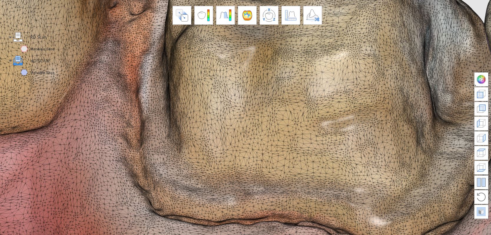
The clash of the titans are in full effect. We are not smart enough to understand all the dynamics of patented software and hardware. While we let the manufacturers sort it out, we wanted to demonstrate how the new mandibular jaw movements are captured in Meditlink
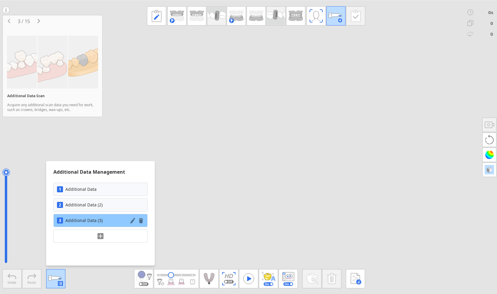
Many scanners limit the number of scans you can take on a patient. This becomes cumbersome when you want to image pre-existing conditions, tissue scans for implantology, and other advanced needs
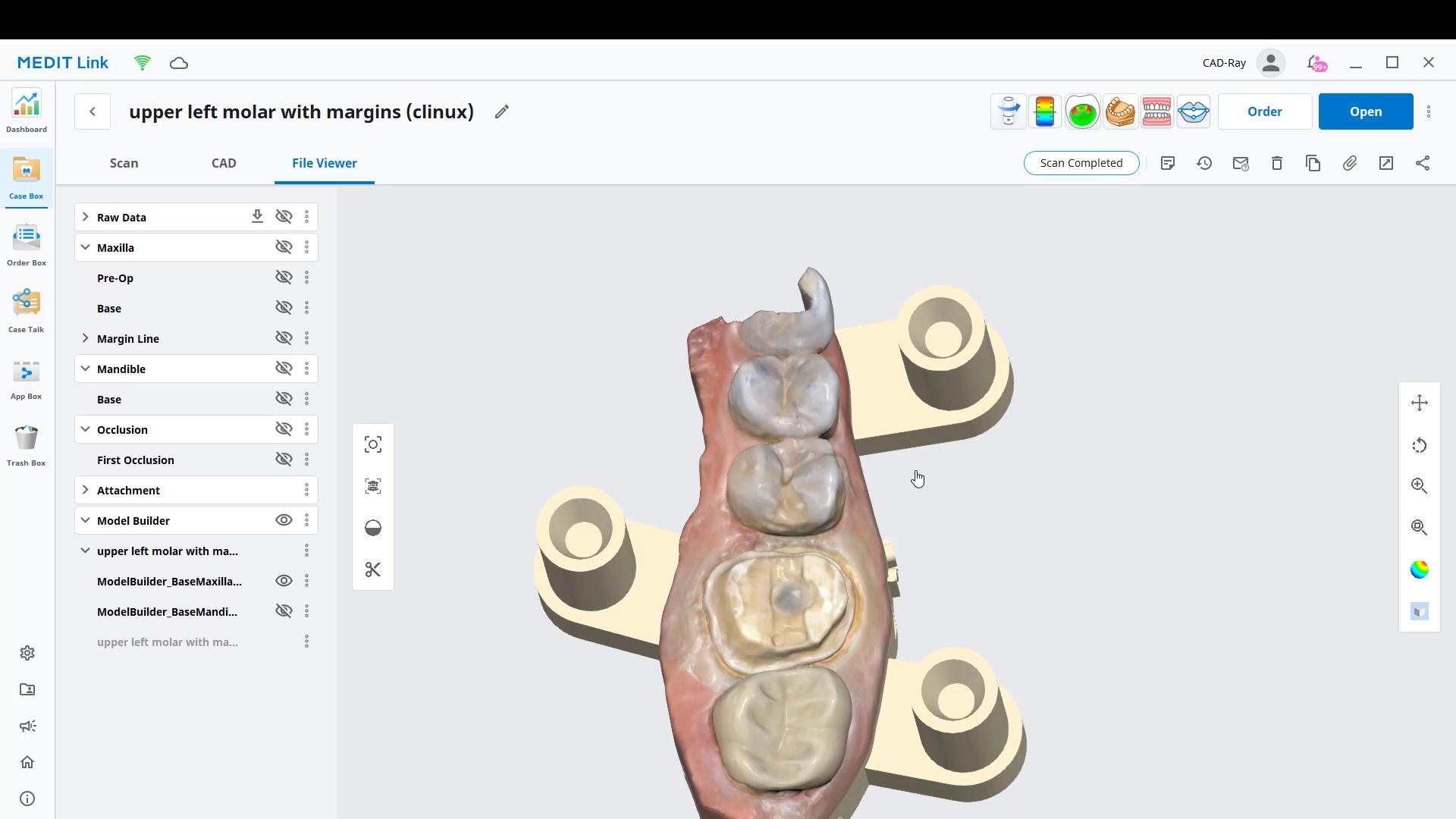
With version 3.0 in Meditlink software, launched in October of 2022, you can import or scan multiple models in the same patient chart
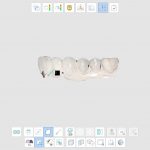
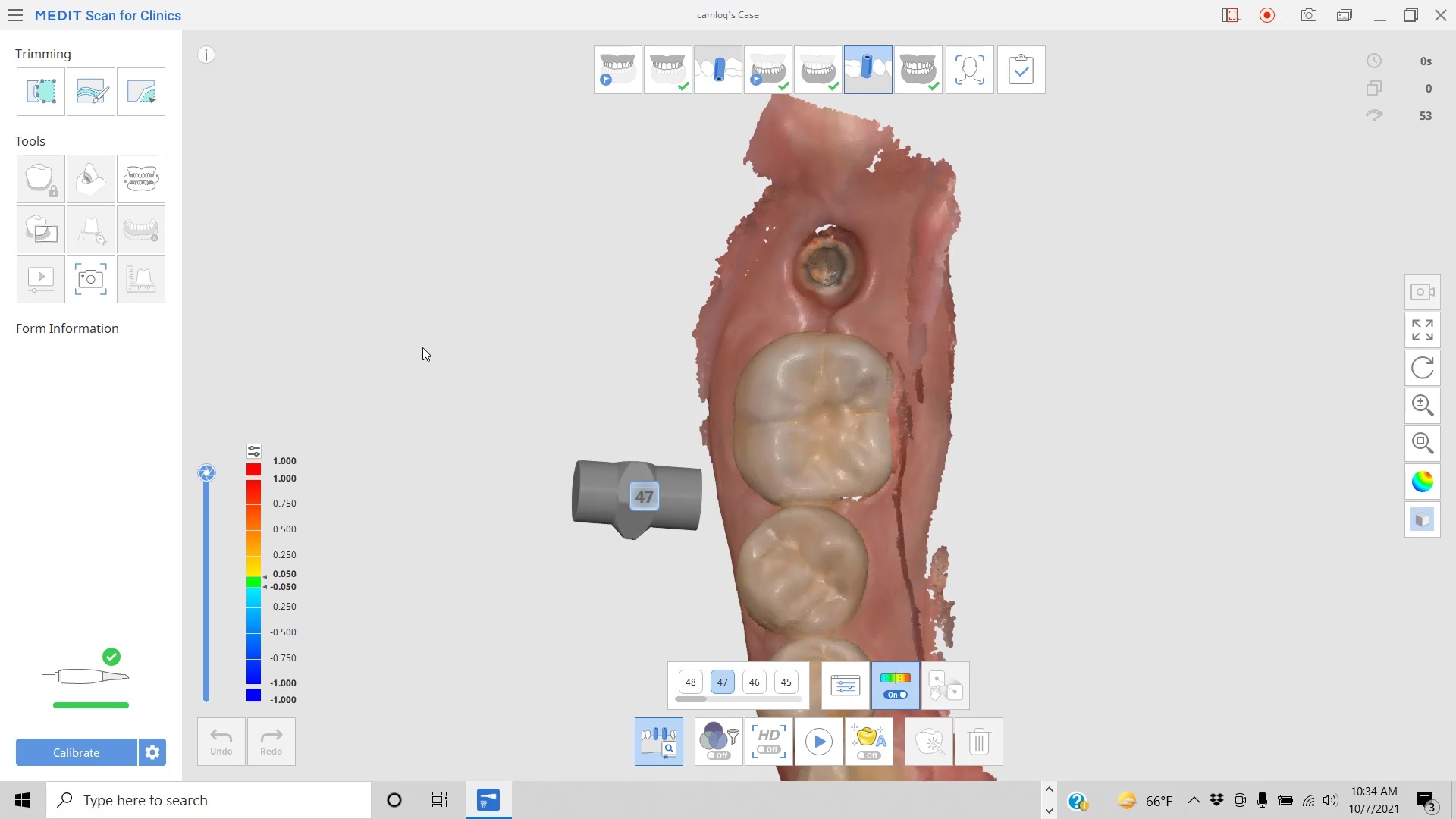
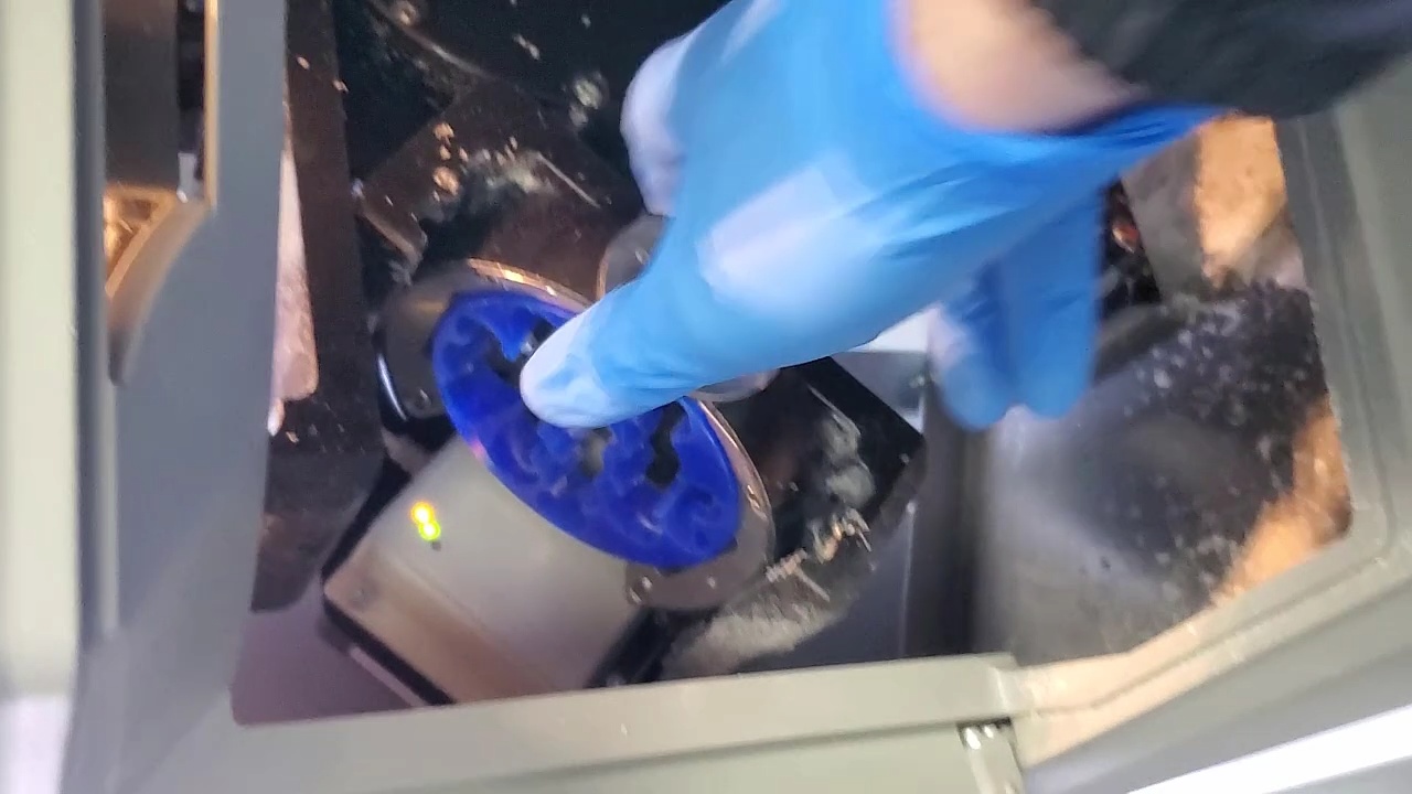
In this presentation we detail the implant suprastructure identification system where an existing abutment is removed and scanned extra-orally to capture the margins readily after the abutment is anodized
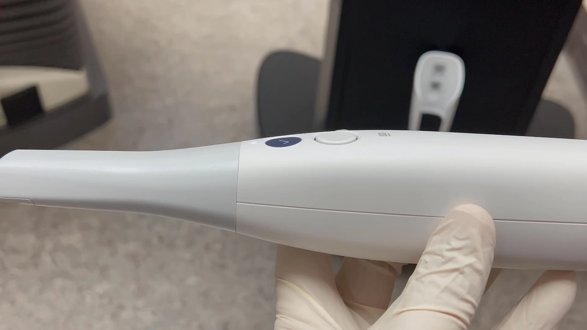
In April of 2022, Medit launched its wireless version of the i700 intra-oral scanner. We’ve been testing it ever since and are impressed with the range that it has. You want to be within 10 meters but more importantly you want to have clear line of site from the scanner to the hub. Much like anything else that is wireless, the further you are from the receiver the more difficult it is to maintain connection. In this video we demonstrate the distance at which you can maintain good connectivity.
After connectivity we tested its ergonomics. Some users get frustrated with tangled cords that are teathered to the laptop computer. This is a great solution and there is not much weight added to the device making it very easy to handle
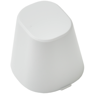
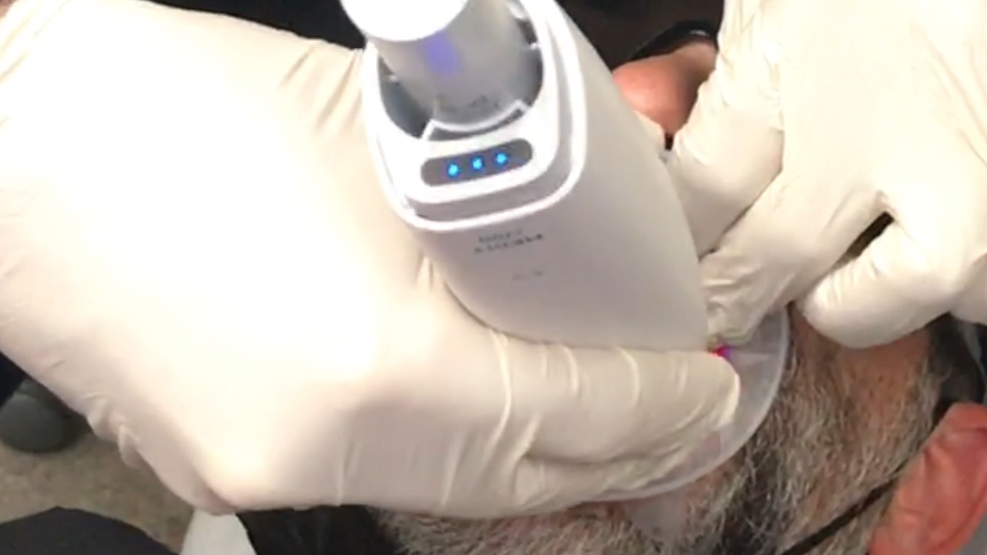
In this video we show the layout of the battery pack, the scanner, and the dimensions of the battery pack. It is very easy to remove them and swap them with fully charged nodes.
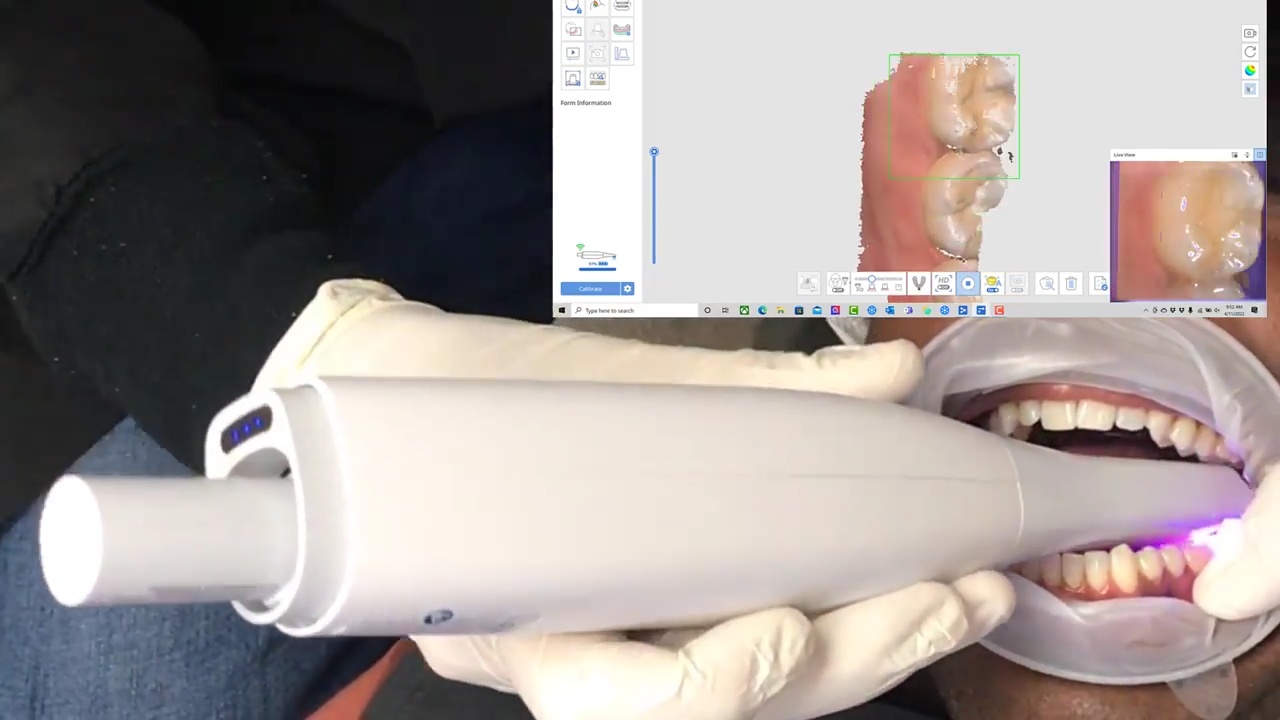
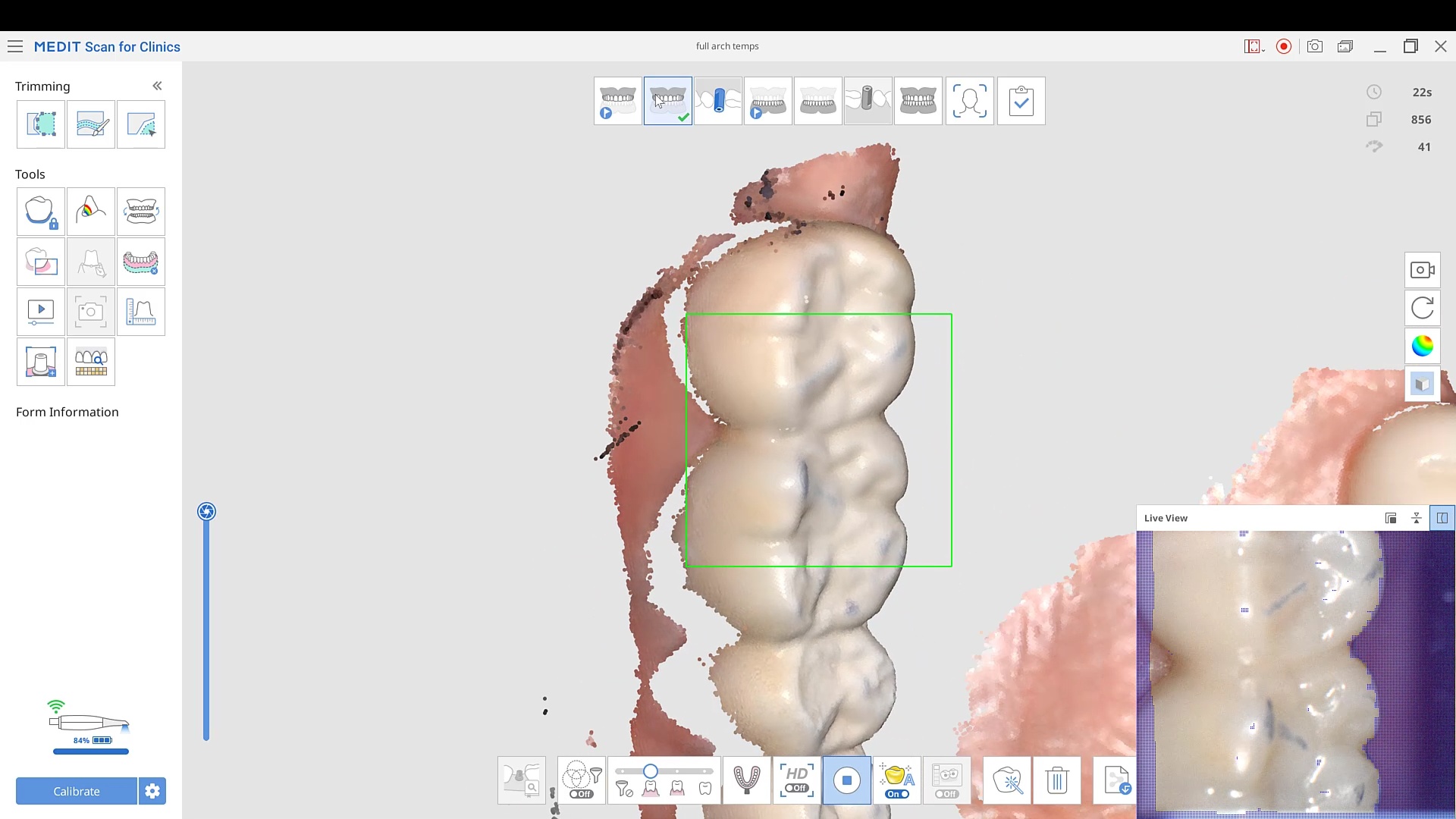
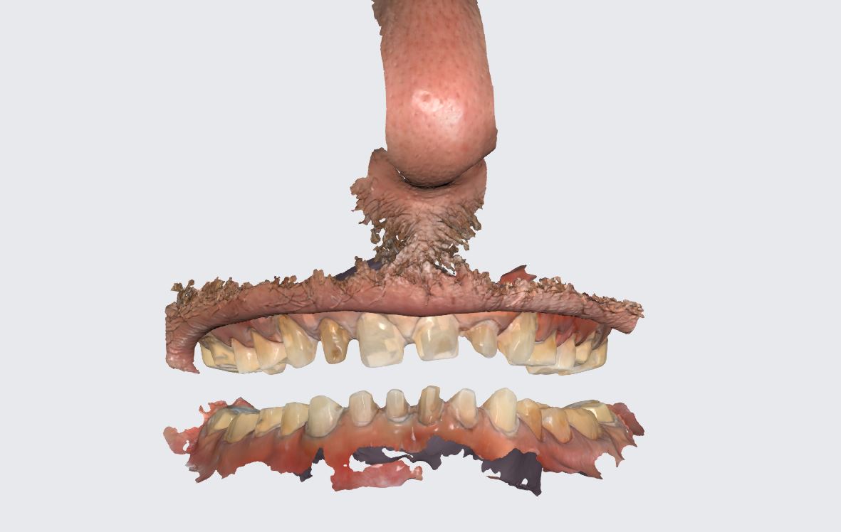
In this video we had the chance to scan a full set of upper and lower temporaries. You can see all the great features of the scanner in action where we capture the maxilla and mandible in little time.
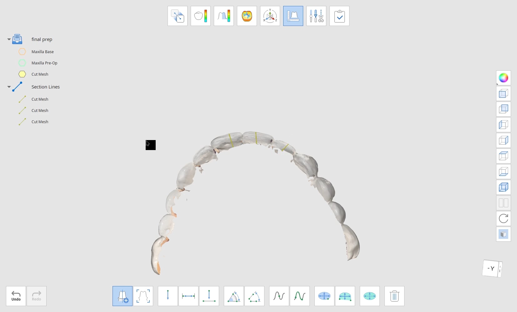
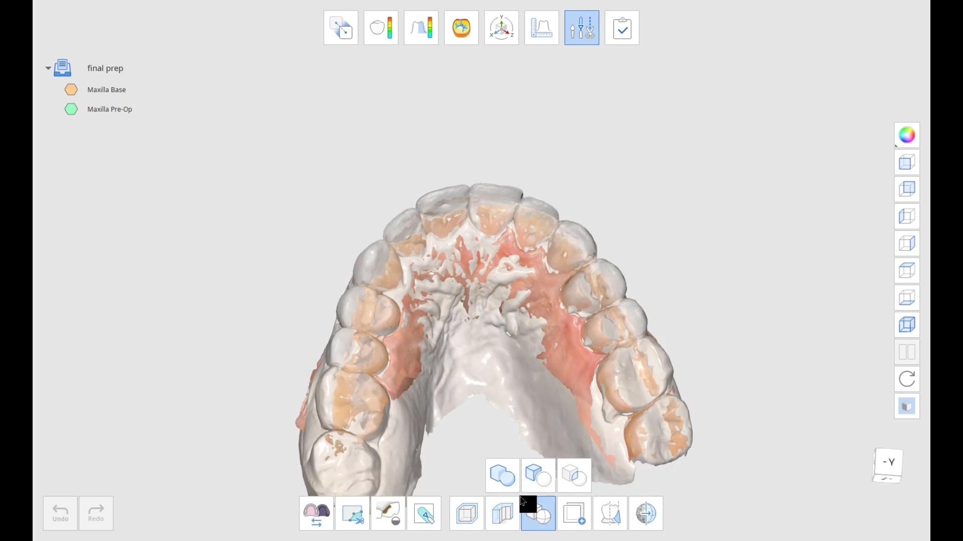
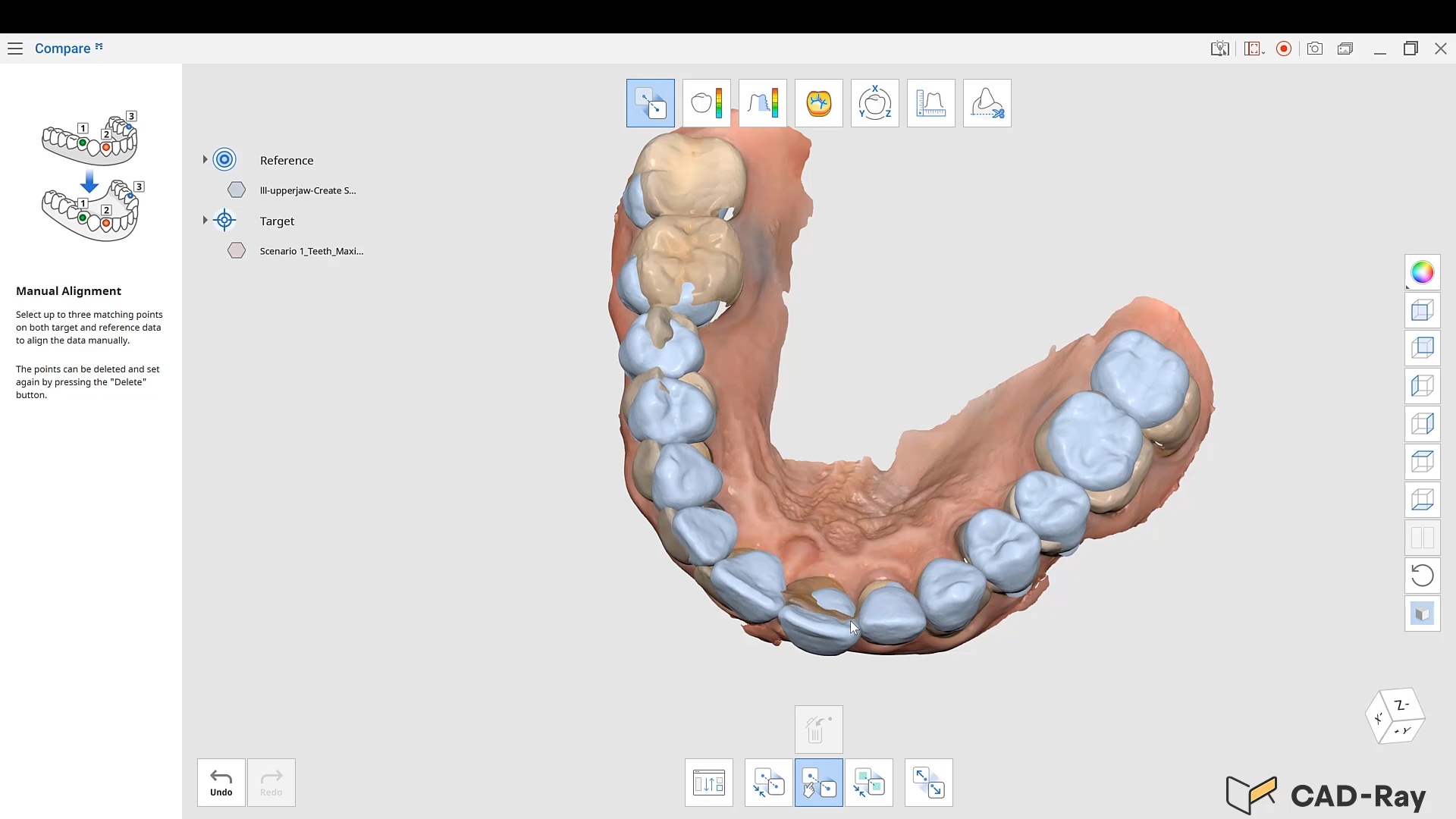
the patient was sedated and intubated for the case so we could not keep track of the bite. Instead, we imaged all 30 prepared teeth and used medit compare / design to digitally mount them to the wax ups. In the link provided you can download the models and relate them to each other […]
To access this page and view the premium content and support, you must either be a customer of CAD-Ray or purchase CAD-Ray Membership.
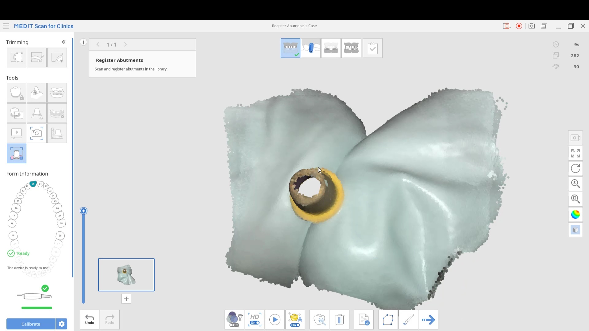
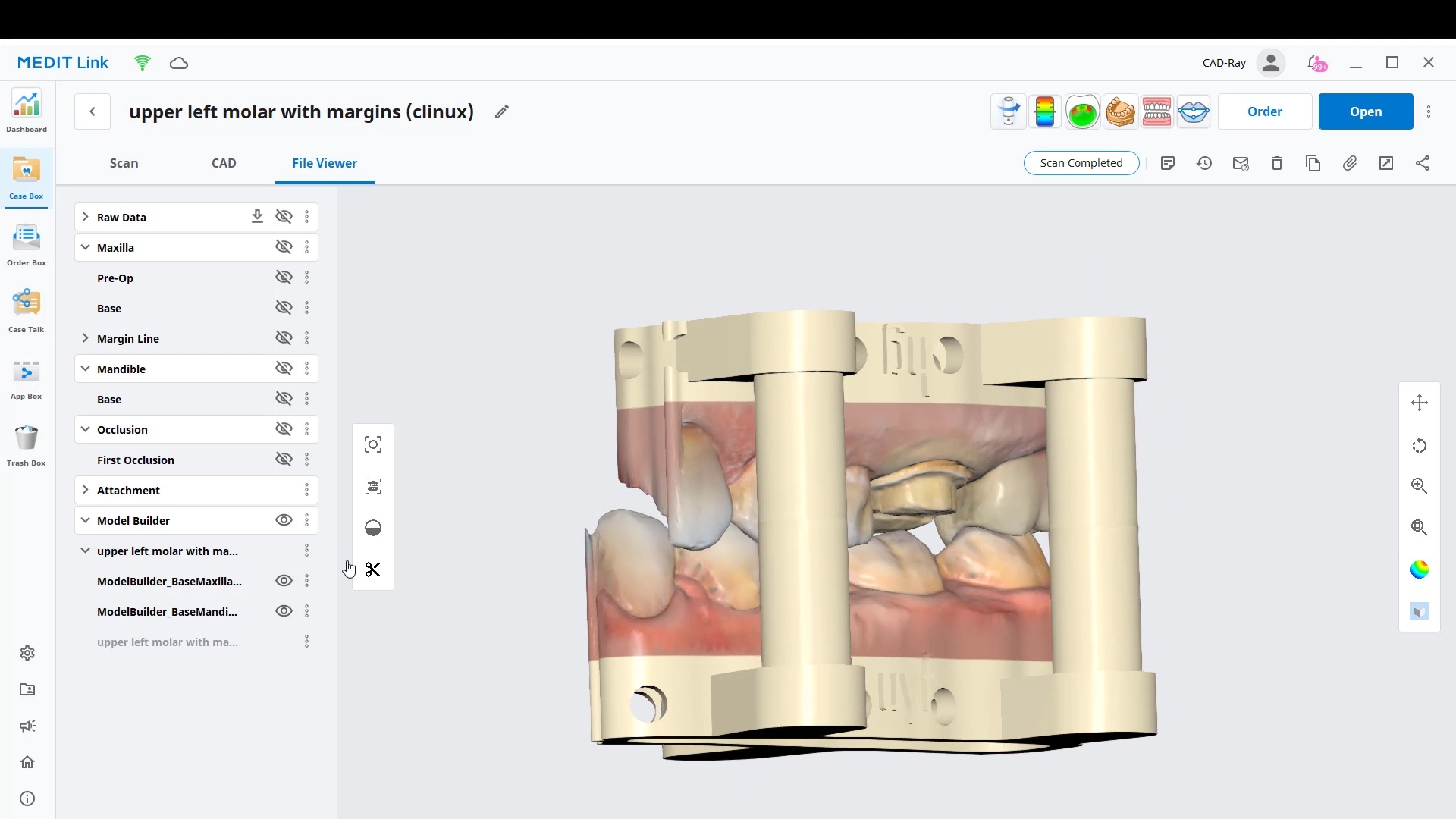
Medit Compare, now called Medit Design, now has a boolean cut feature that lets you extract a temporary shell model from wax up or mock up model and prep model for easy and quick designs without painful margin marking on multiple units
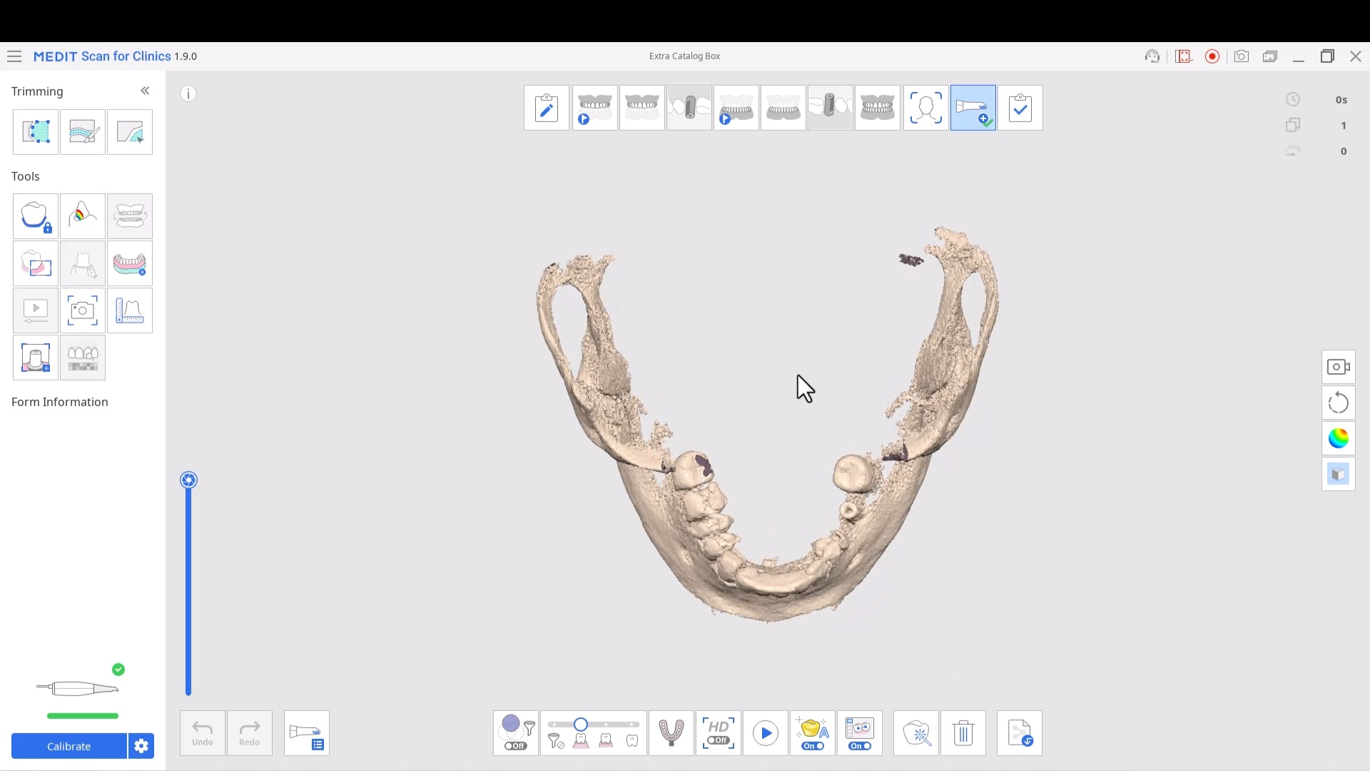
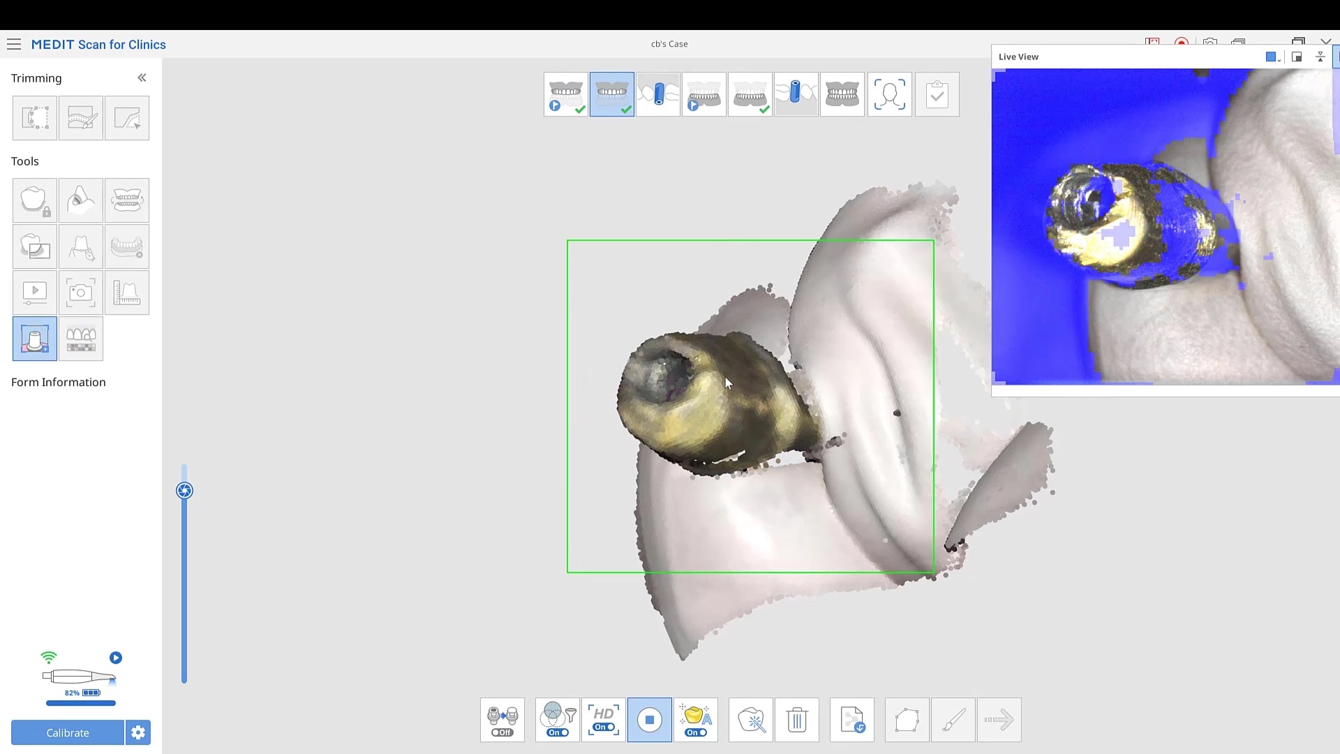
Medit’s software allows you to remove and pre-existing abutment and scan it outside the mouth. This allows you to find your margins without ever having to displace the tissue or reach hemostasis. Important matters to keep in mind with workflow
You must fill out the Rx form correctly and identify the abutment location
You must take the abutment scan at first in the right catalog box (upper jaw or lower jaw)
You must take the tibase / abutment outside the mouth and scan it under Abutment Registration Feature
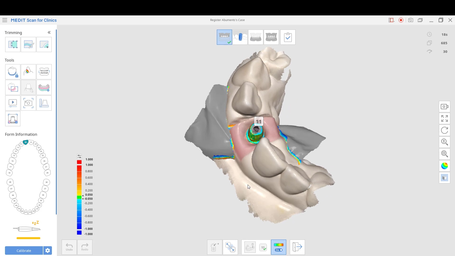
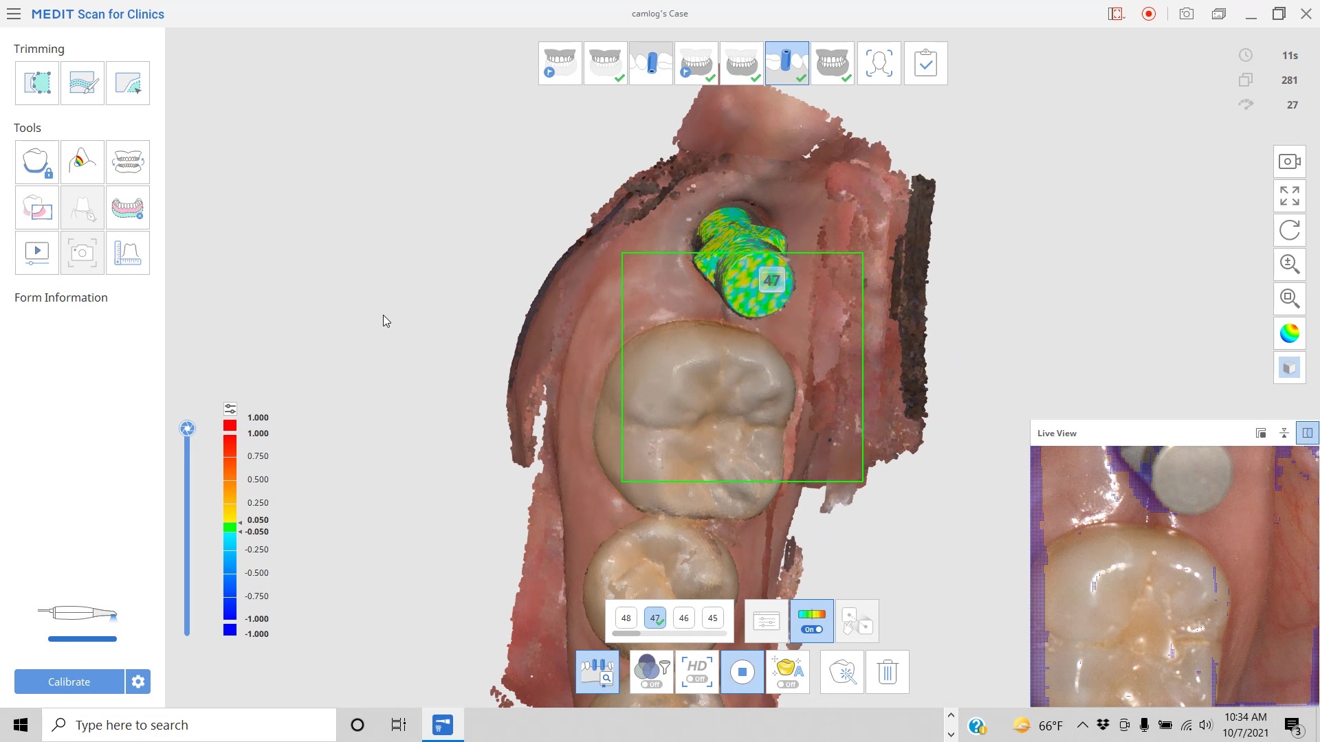
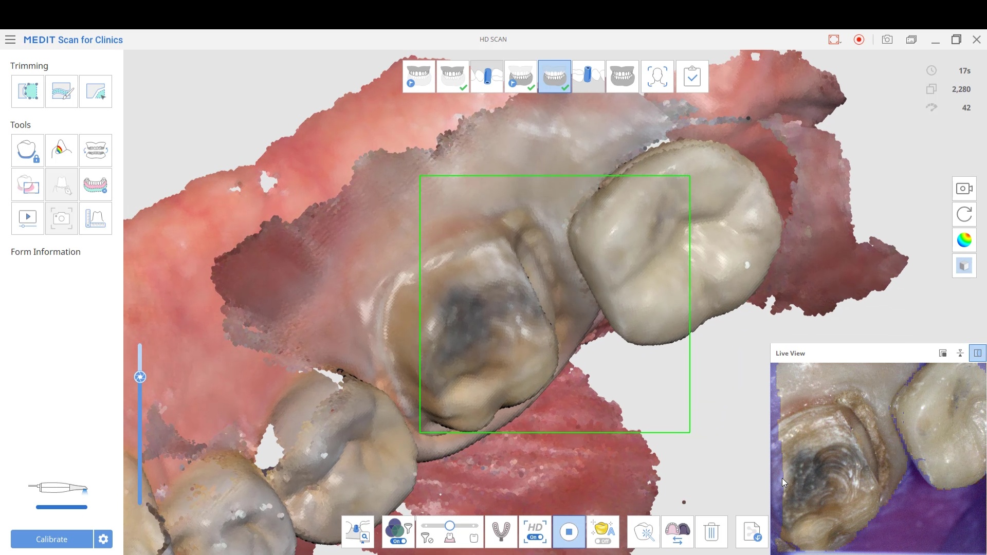
In this video, we show how you to capture the healing abutment, the tissue profile after the abutment is removed, and then we image the scanbody while utilizing the AI feature of the Medit i700 to pick up all the data from the scanbody by matching the STL to the physical one during the scan
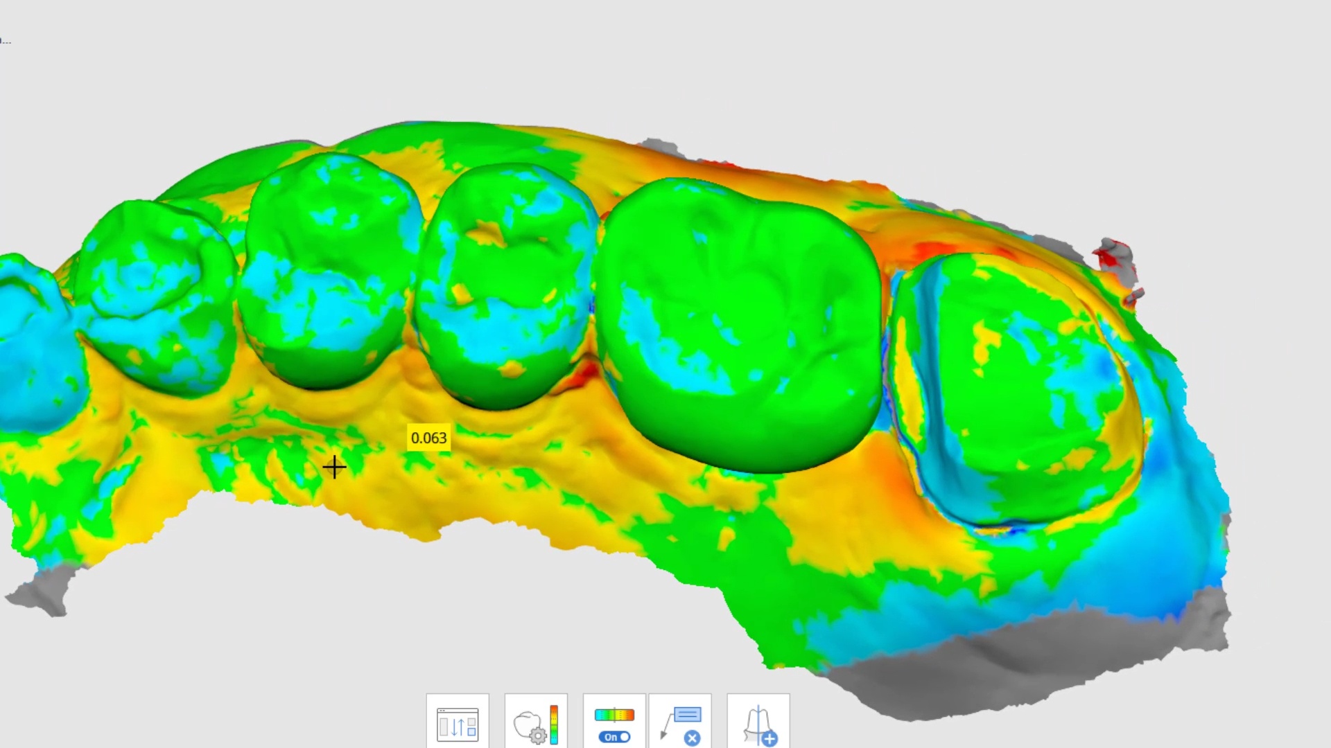
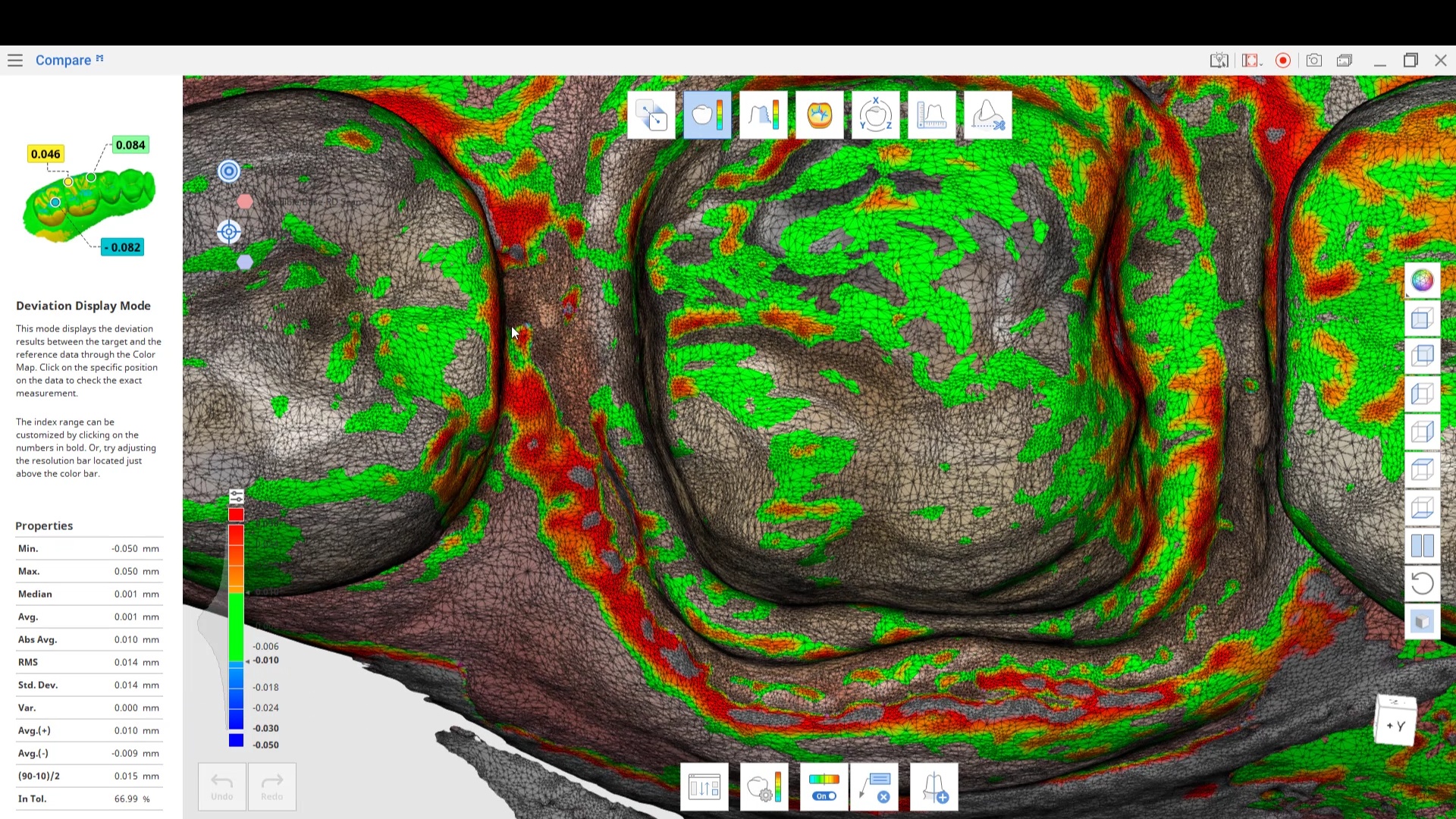
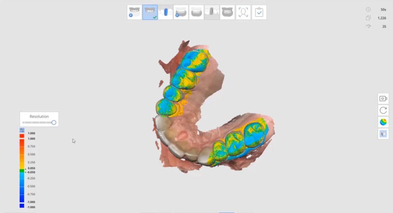
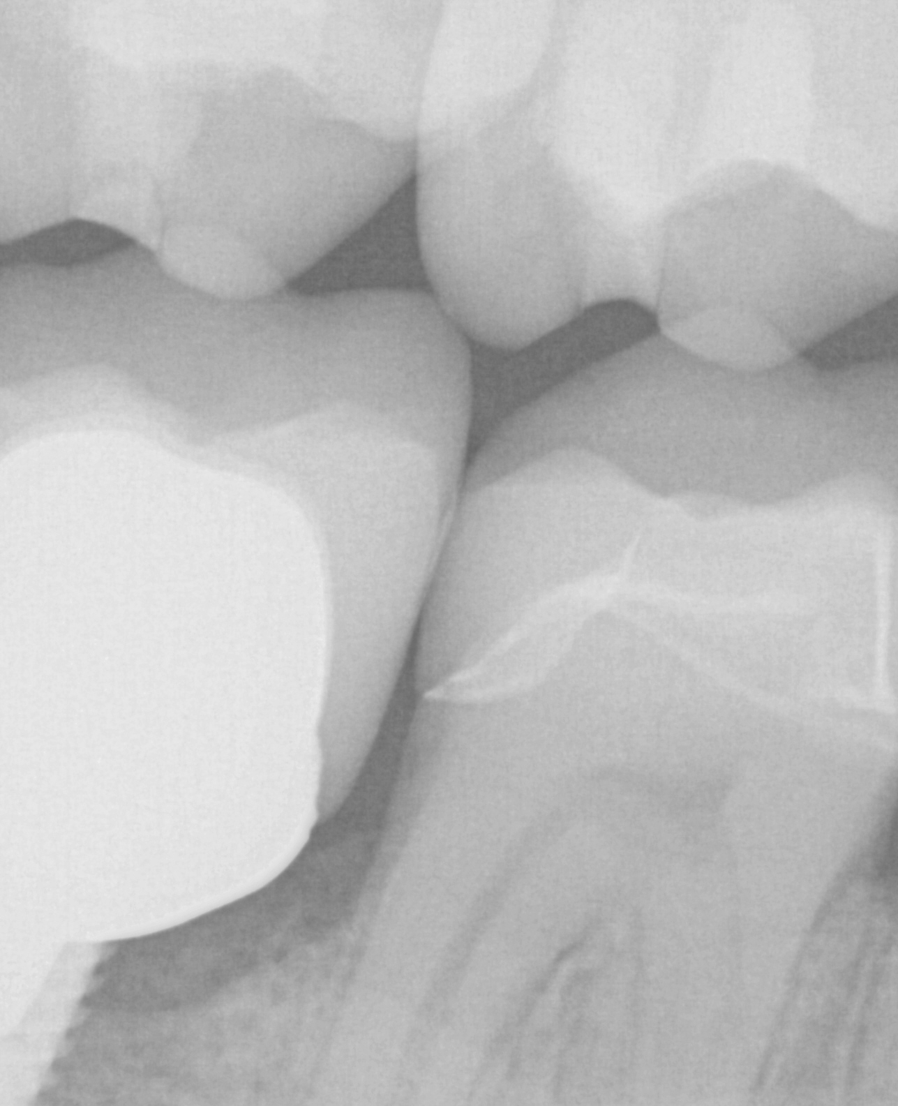
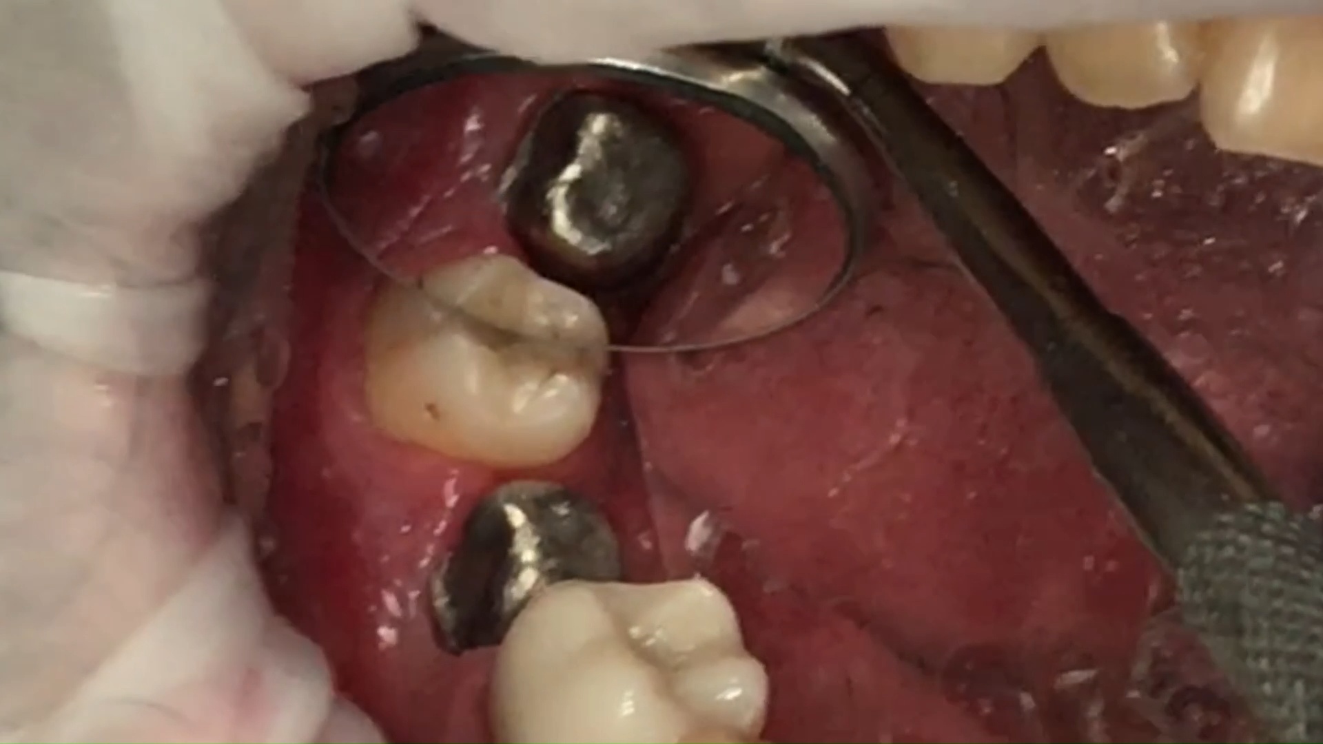
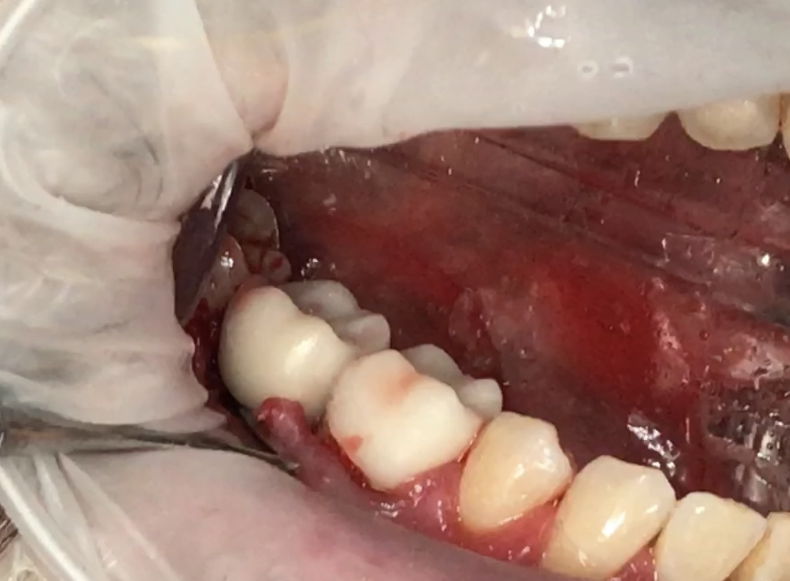
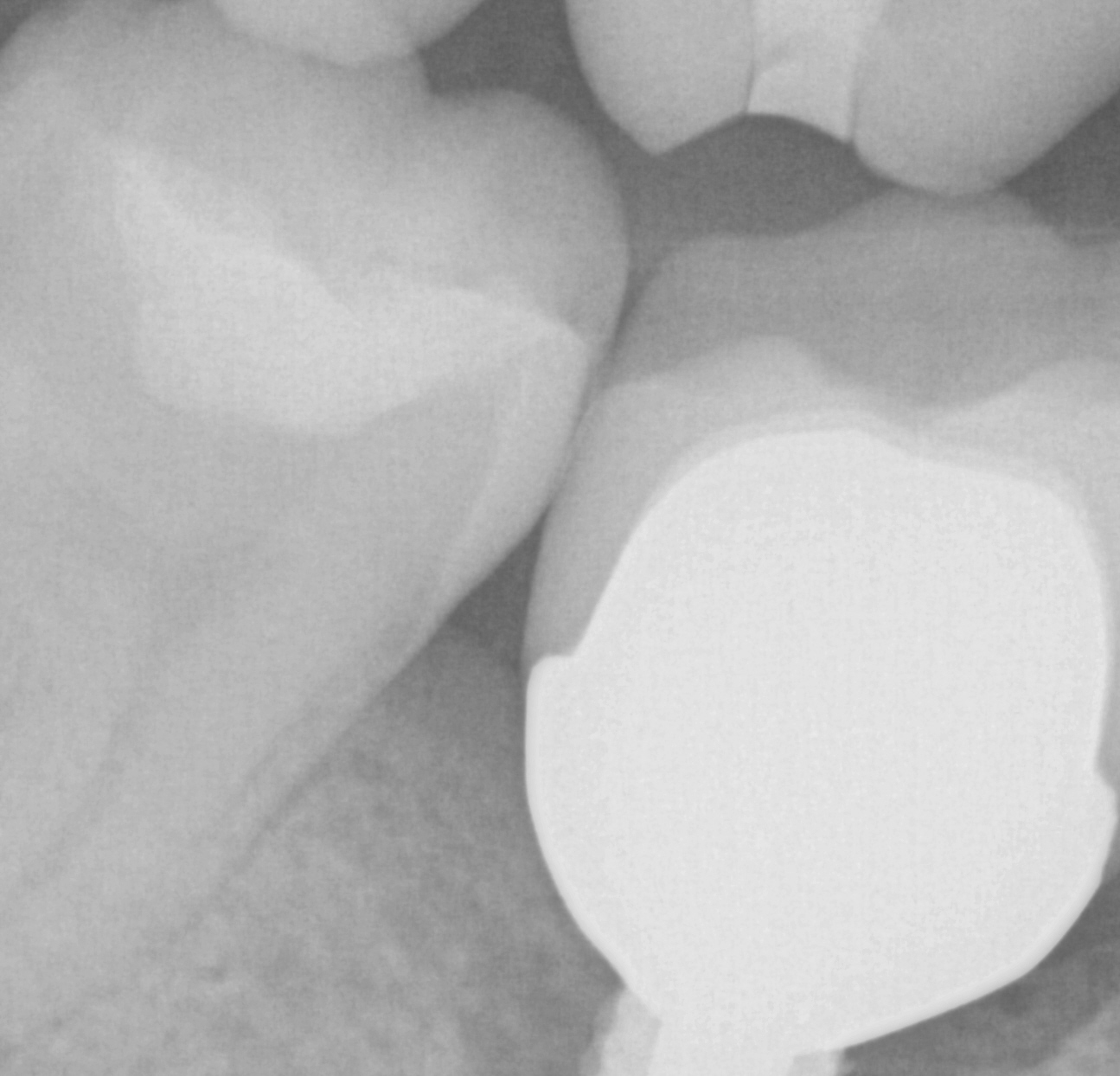
With second molars, you should always be on the look out for not just the jaw settling, if you remove the first point of contact, but also with the temporary step forcing the tooth to tip towards the distal, if there is no third molar to stop its tilting.
In this case, a doctor was trying to seat a second molar crown he had just prepped a few weeks prior. There was an open contact and he could not ascertain the reason for this. He did take a second impression digitally so we had the chance to merge the two models and look for discreptancies between the preps. This video shows how the comparison of those two steps in Medit Compare
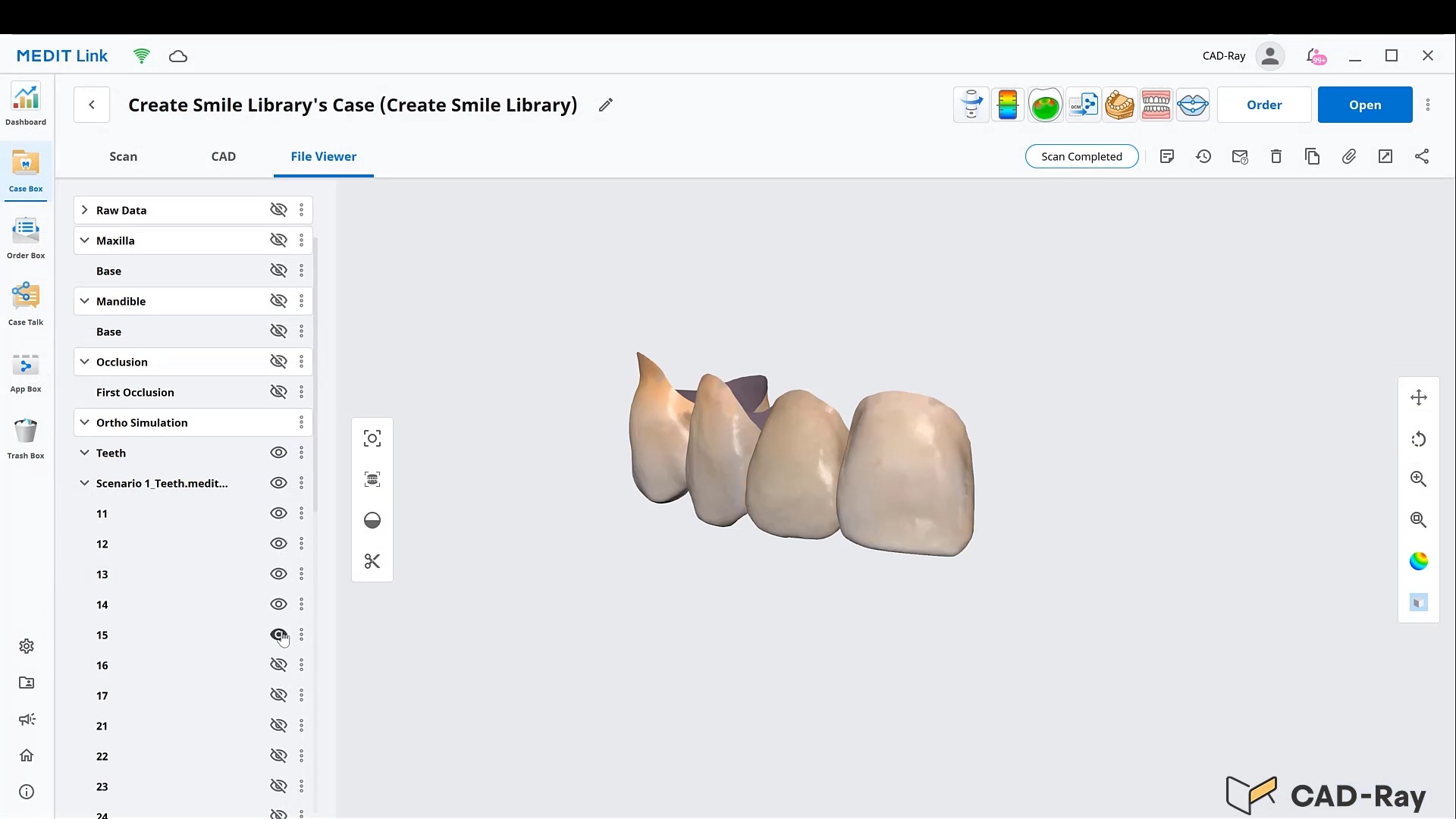
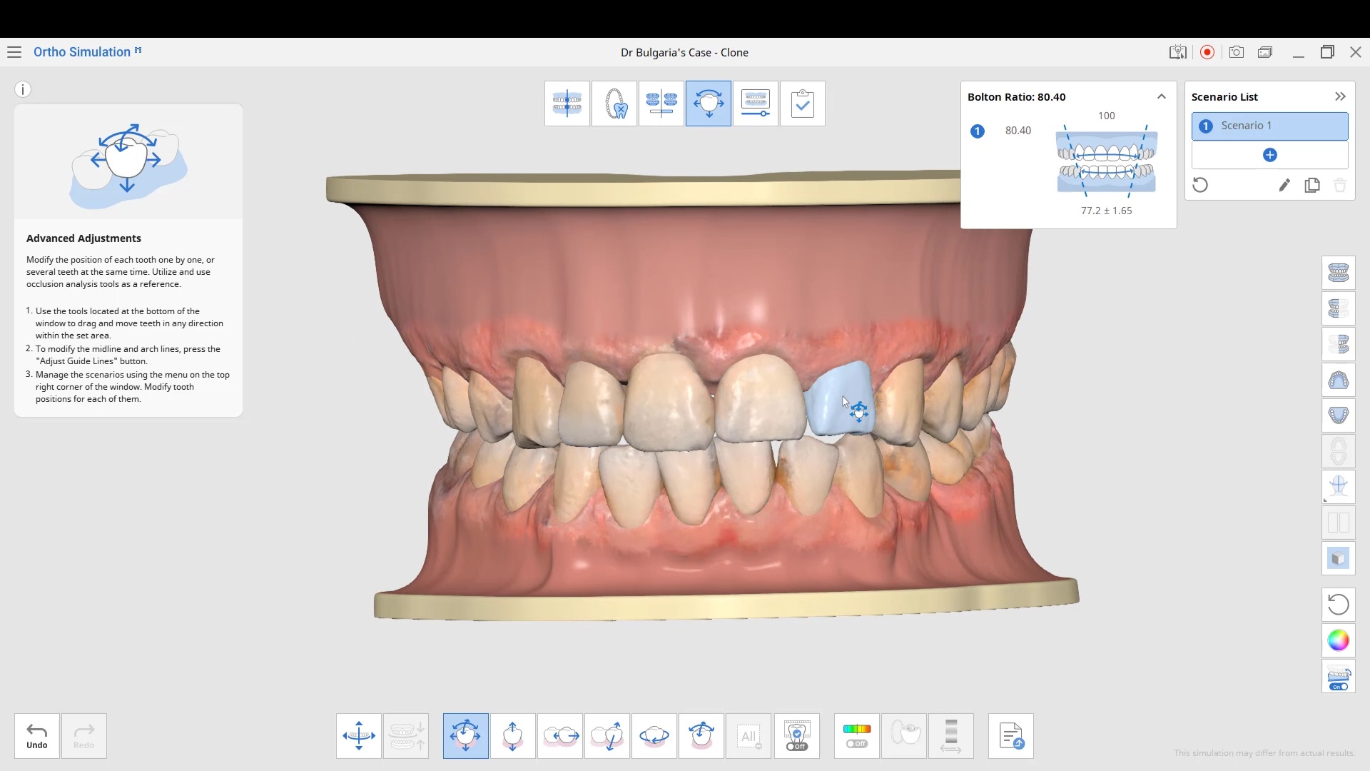
Medit Orthodontic Simulation application allows you to segment out individual teeth from patient scans into an stl format that you can use as a template for smile design wax-ups. You have to watch the vide for it to make sense
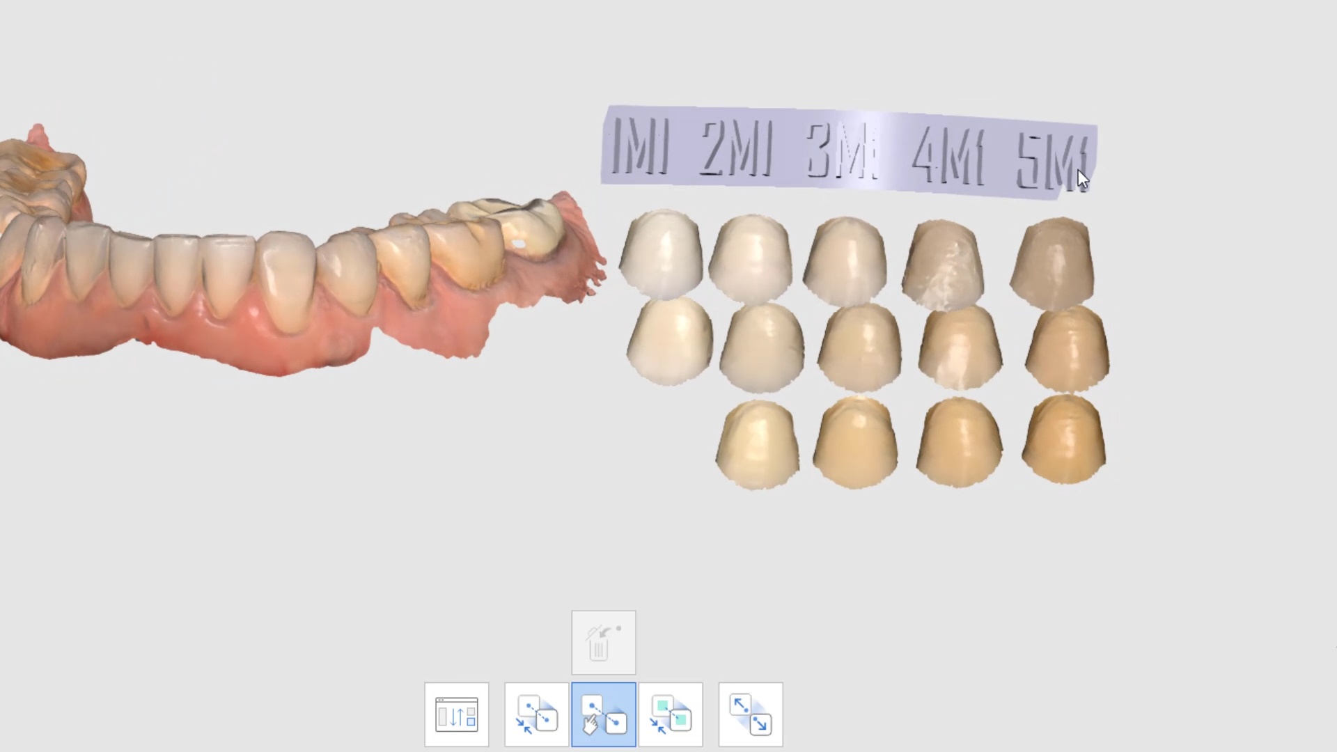
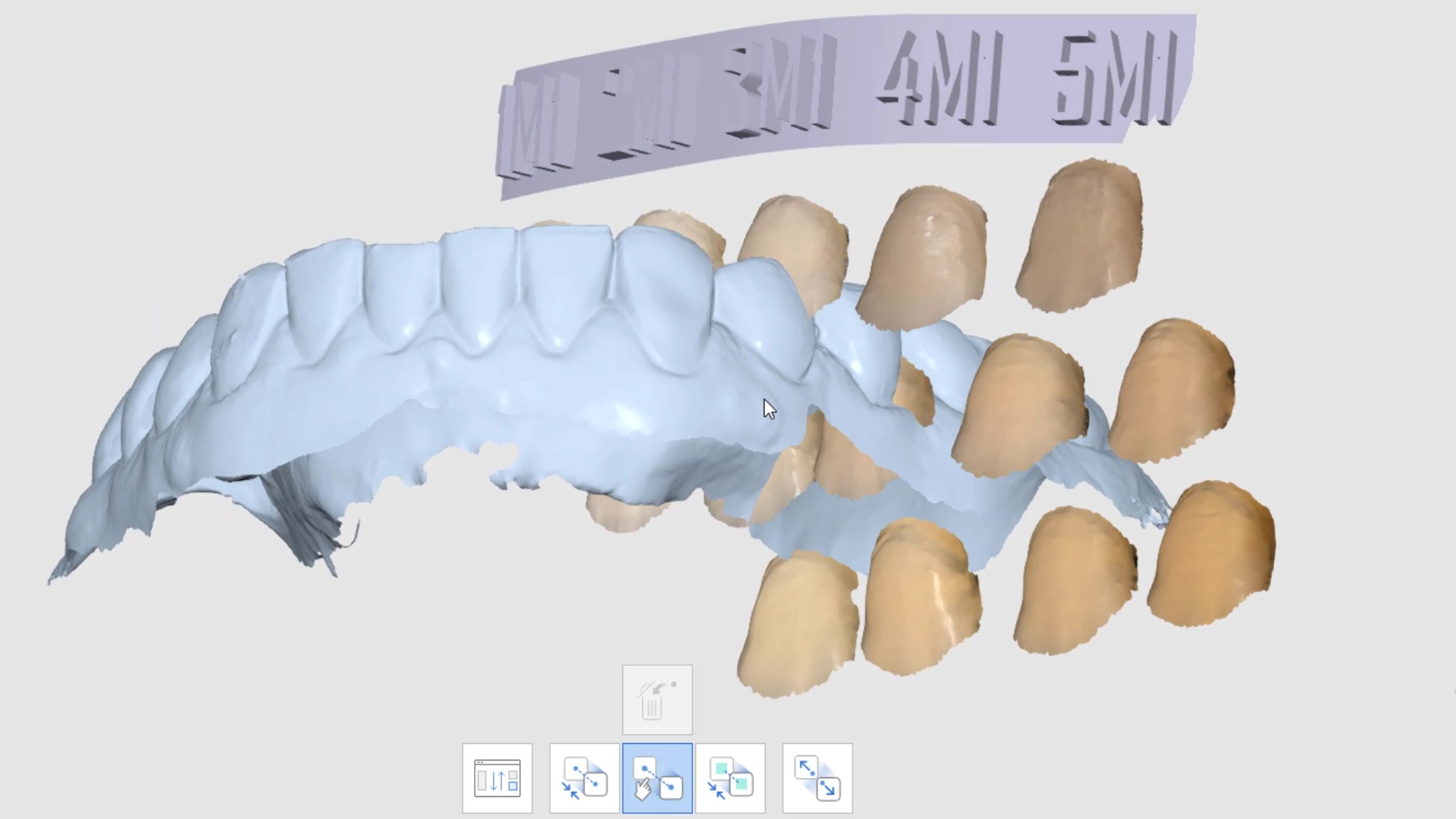
We’ve digitized the Vita 3D shade guide to help with color identification. The same Medit scanner was used to scan the tabs and then this digital file was created. the assumption is that the same camera and light will hit natural dentition and the net effect will be the same. Use at your own risk.
Still not a single article published that says Medit ios is a good scanner ! its just been user driven for 3 years now.
it’s a good thing, because the world just changed. it is irresponsible to extrapolate research done outside the mouth on stone models or impressions into clinical significance with intra-oral scanning. There are parameters that are impossible to quantify like focal distance throughout the scan (unlike desktop scanners with known focal distances), the codes use to do the algorithms, the scan patterns, and also how light is treated by enamel, dentin, and restored materials.
i can’t believe people still use terms like trueness and accuracy when they really don’t even exist when you scan intra-orally. Like analog impressions, it is impossible to judge digital impression accuracy LIVE while it is happening.
Enter Medit! There are a few distinct ways to demonstrate an accurate scan live while it is happening. One way is to import a geometric shape that doesn’t alter its form while models are being rendered. That’s what’s demonstrated here. To my knowledge no one has ever studied this approach because no other camera lets you do this. i did see some publications where the authors attached objects like radiographic markers and after it was processed, they could measure that object and see if it distorted or not, but nothing at this level.
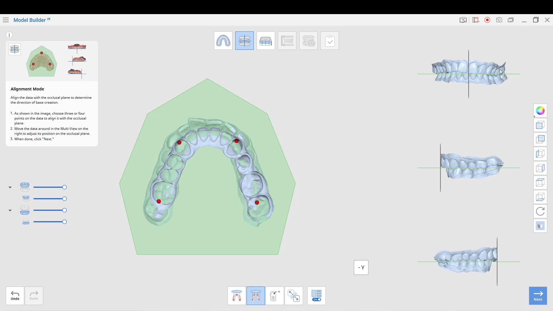
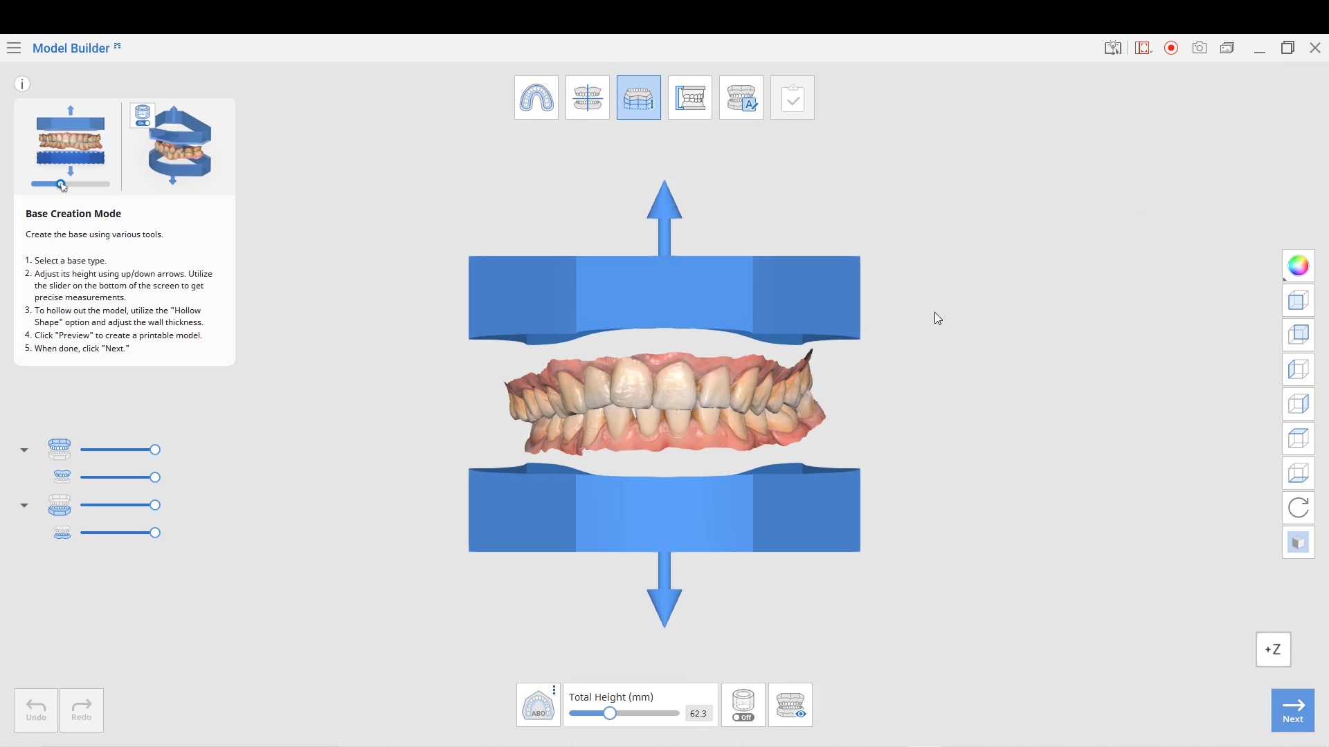
The following information is from the The American Board of Orthodontics. We preview how the Medit Ortho Simulation and Model Builder can satisfy their criteria
Medit has many free applications, including Smile Analysis, Orthodontic Simulation, and now, Medit Model Builder
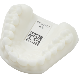
Adding bases has been a feature for a while but as usual, Medit was quick to respond to its users’ request by expanding the base to including holes for drainage in printing and by allowing us to add support pins to the models. Many people who have printers, whether labs or clinicians, easily lose the proper vertical relationship when they print the models. By adding these pins, it allows you to index the models properly for any finishing work someone may need to do.
Advanced users are unlikely to use this feature for a single unit, but it comes in handy for oral appliances, particularly when they have advanced the jaw to open the airway. Oh, and this app is free and you can use it with models created by any scanner
Here is why the Medit i700 is the best intra-oral scanner on the market if you do any kind of implant restorations. There are so many options and tools that are leap years ahead of other scanners and their software. Medit can automatically identify the scanbody for you so you don’t have to do cartwheels and gymnastics to pick up all of the scanbody. This is in part 1 of the video.
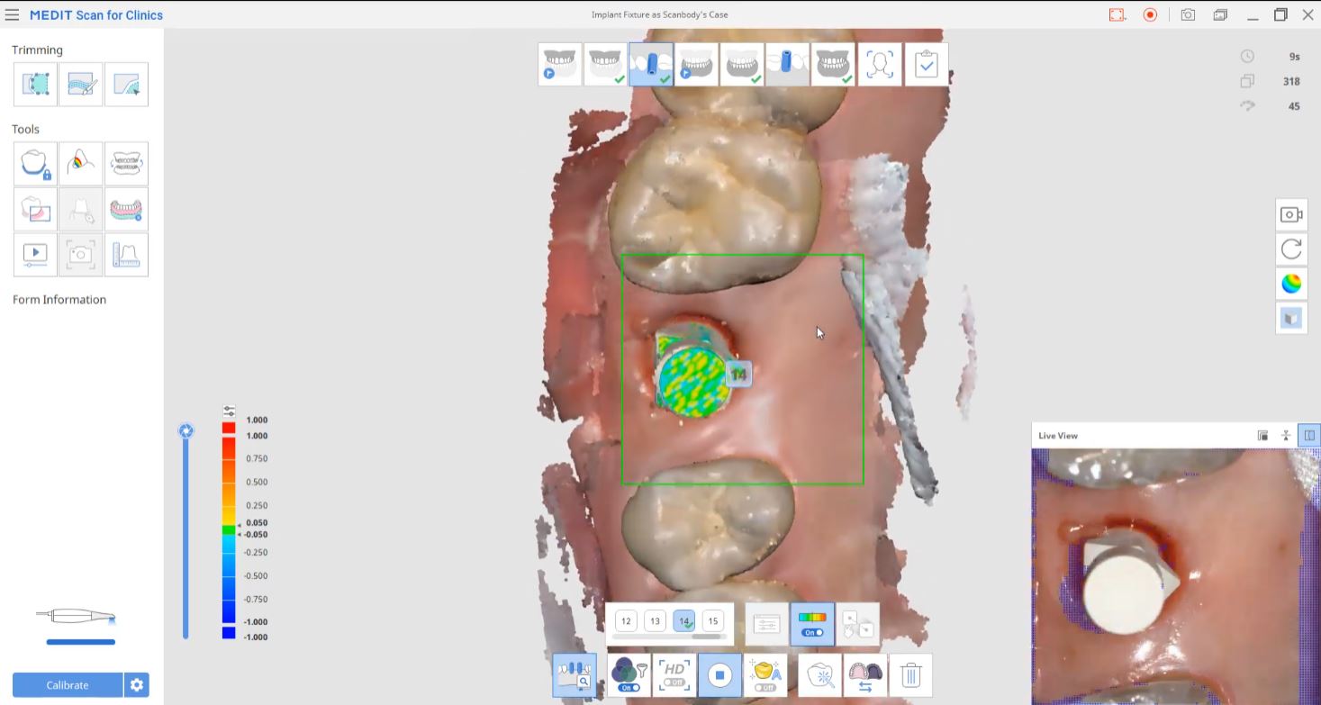
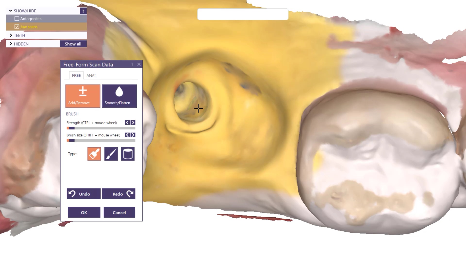
For advanced users, we are sick of dealing with scanbodies and checking to make sure they are seated all the way and not binding on the tissue or bone, so we developed this technique of just scanning the fixture itself. It is not ideal just yet, but it will be the future, as the inside of the fixture is too shiny. i just used some old cerec spray to mask the topography for this demonstration.
oh, and really, no one else can show you how to milk that medit like cad-ray.com can. we use it well beyond what it was intended for and frankly you are wasting your time and money with most others. contact Frank DeLuca, Frank Weinstein, Laura Geney, Nick Statly, Damien Bonner, Jonathan Acker in the US or Milos Gedosev, Mariangela Di Nato, Roddy MacLeod in Europe for more information
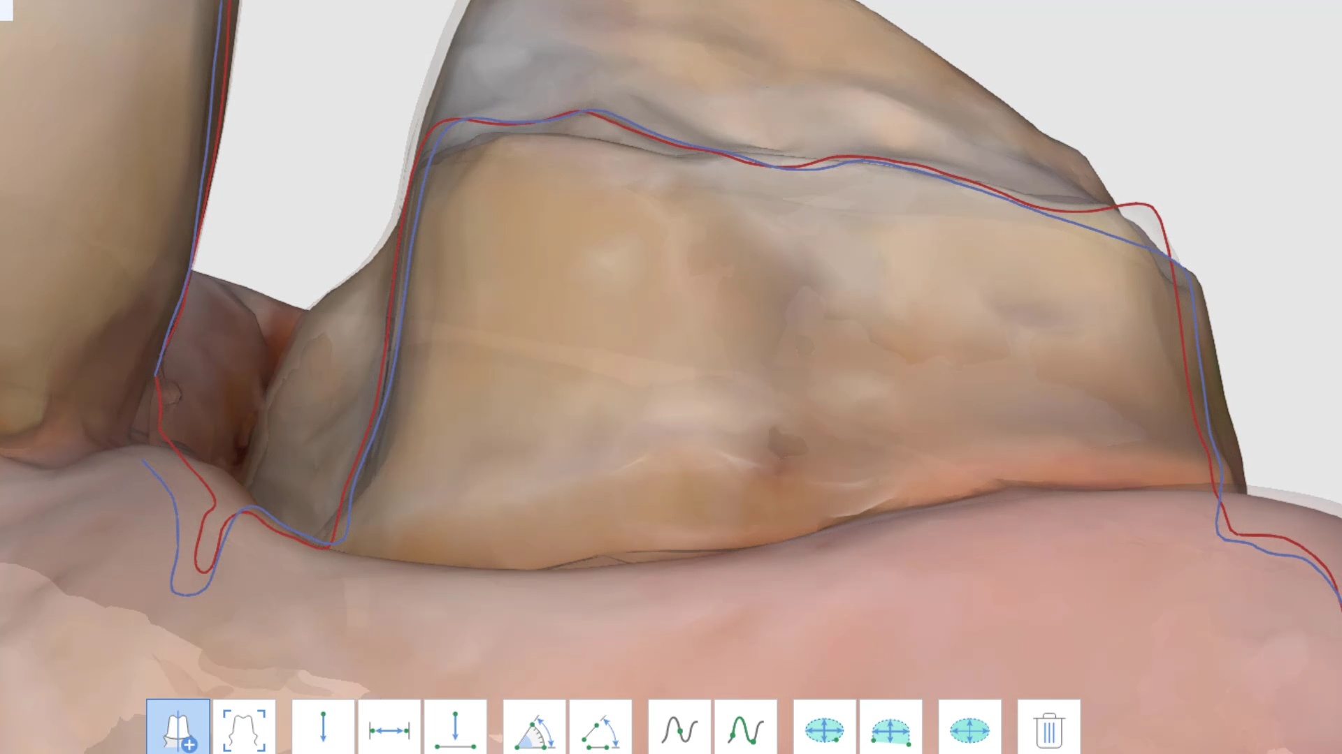
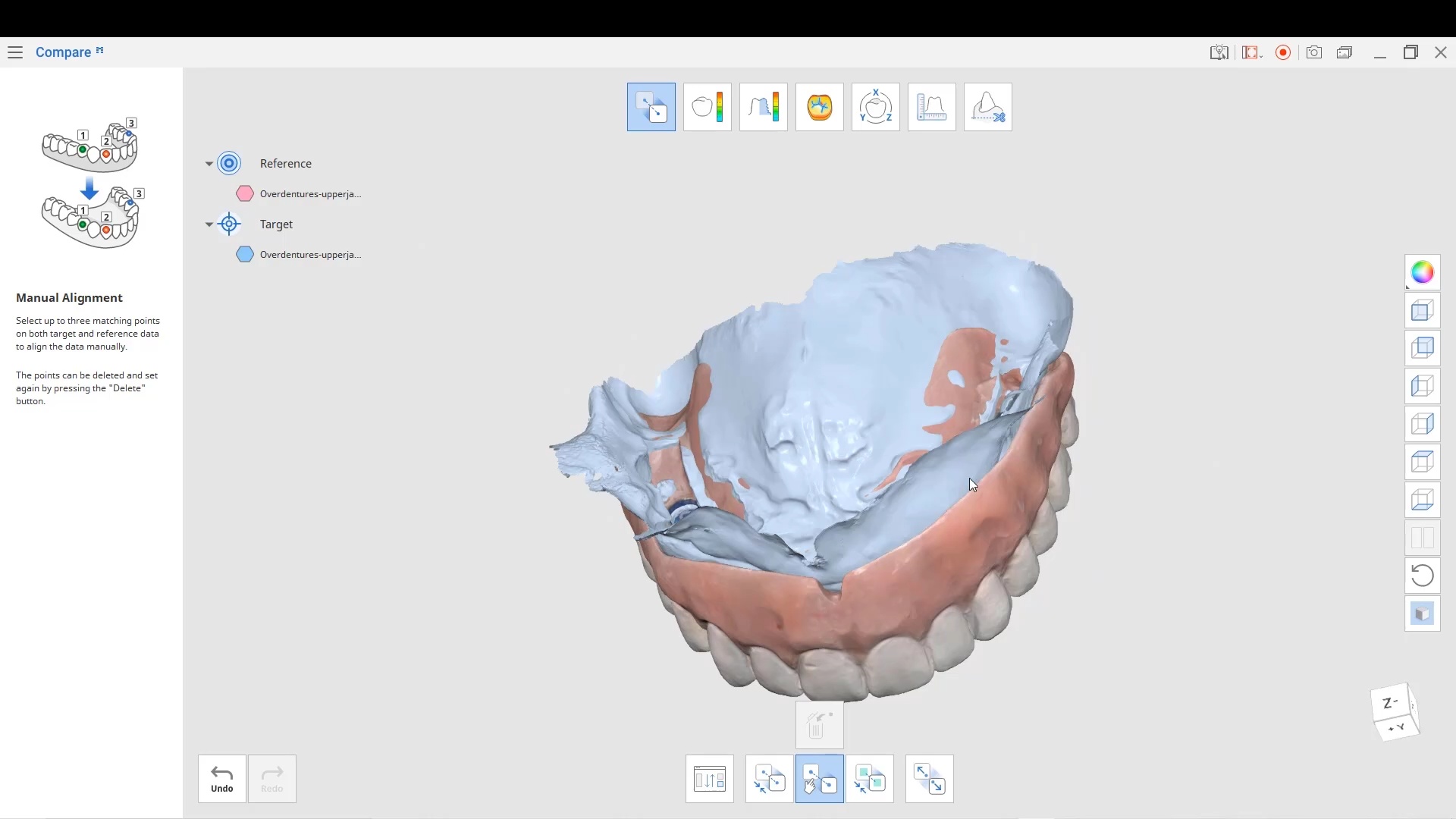
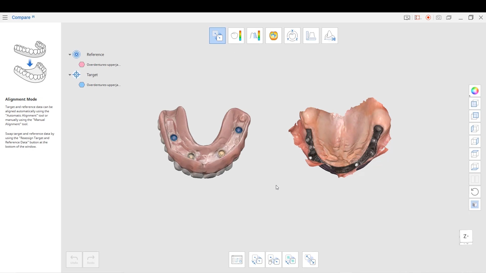
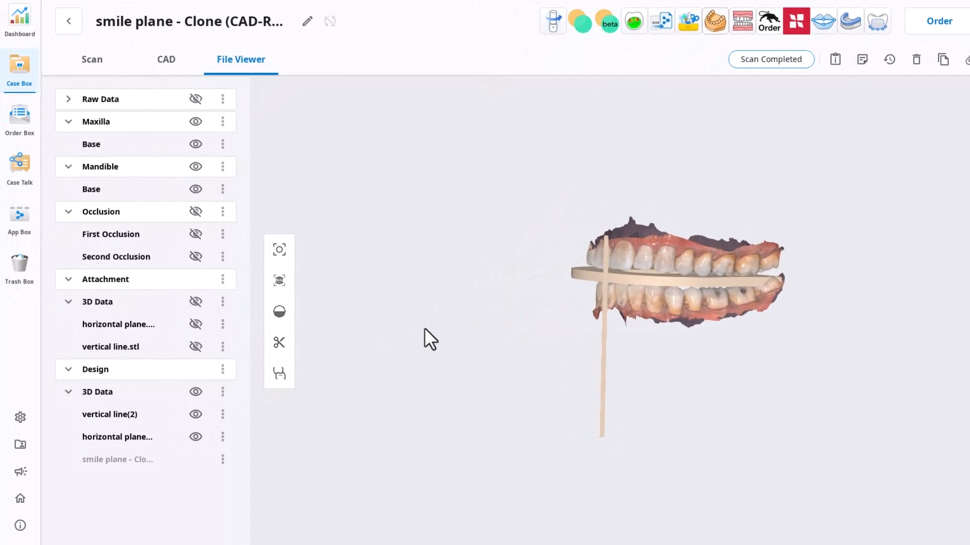
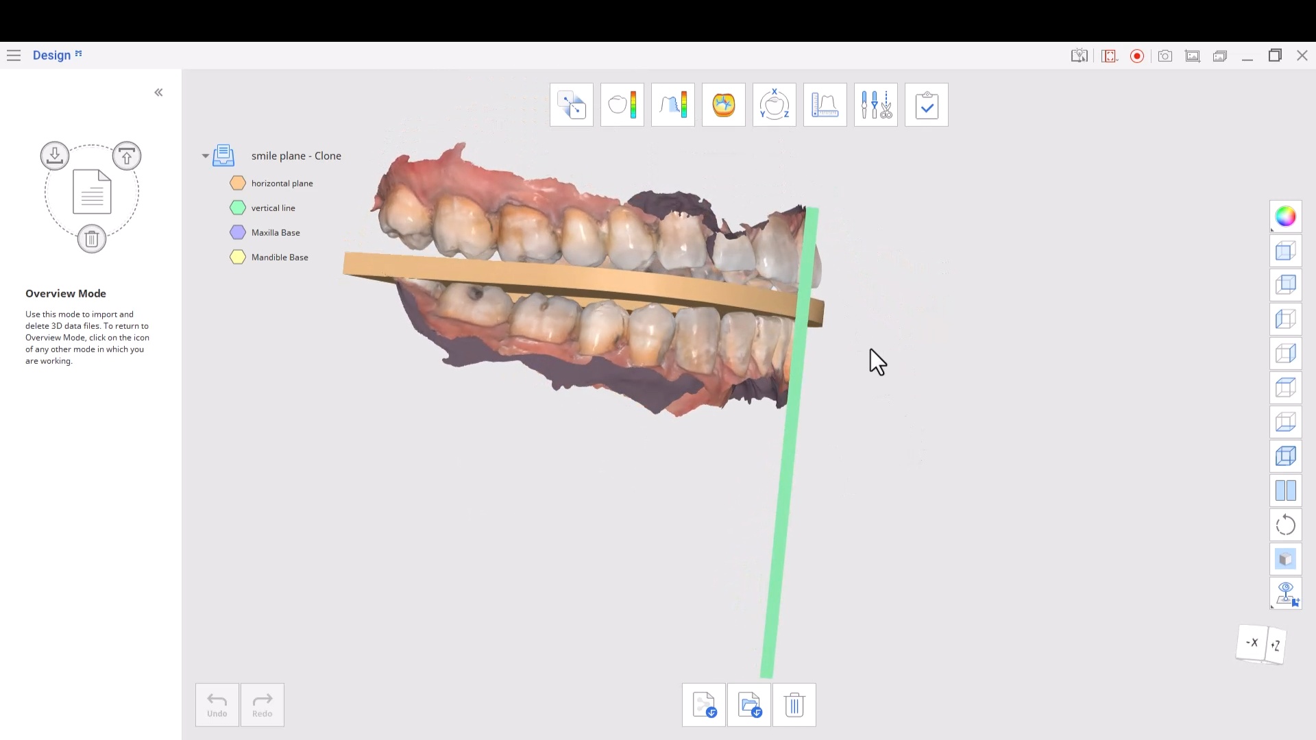
This case shows how to relate models to each and maintain their relationships. It features the power of the Medit Compare app that lets you duplicate models, trim them, modify them to your liking. There are some very useful features in this app that can come in very handy
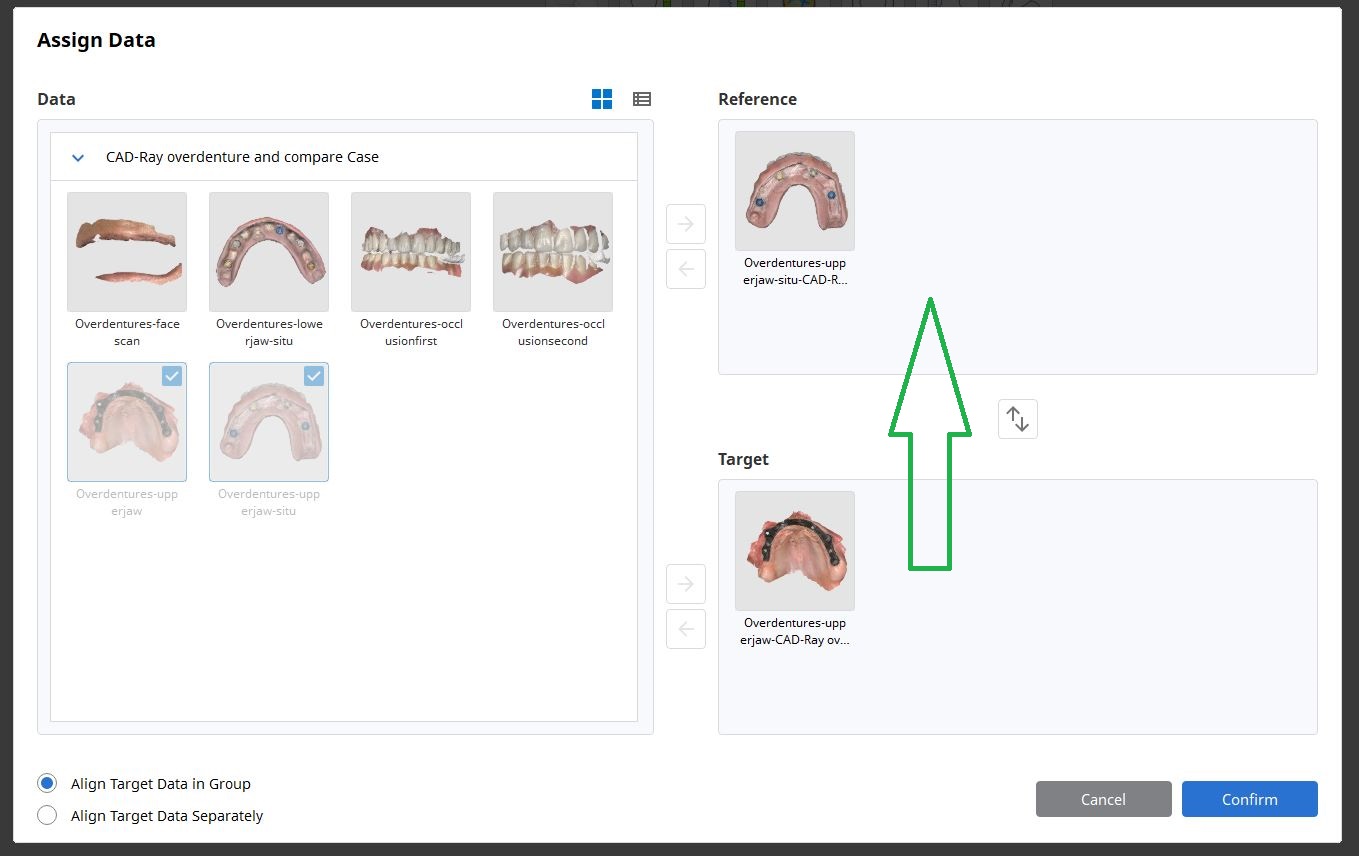
an important matter to remember is the “direction of travel” which is taking models from the Target folder and moving it to relate it to the Reference folder.
All models placed in each folder will maintain their relationships to each other when traveling to connect with the other models. Inverting models will also come in handy for advanced users. You can see the full video and download the case and design along here
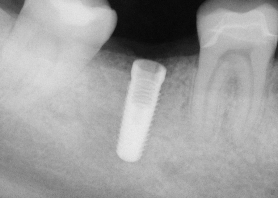
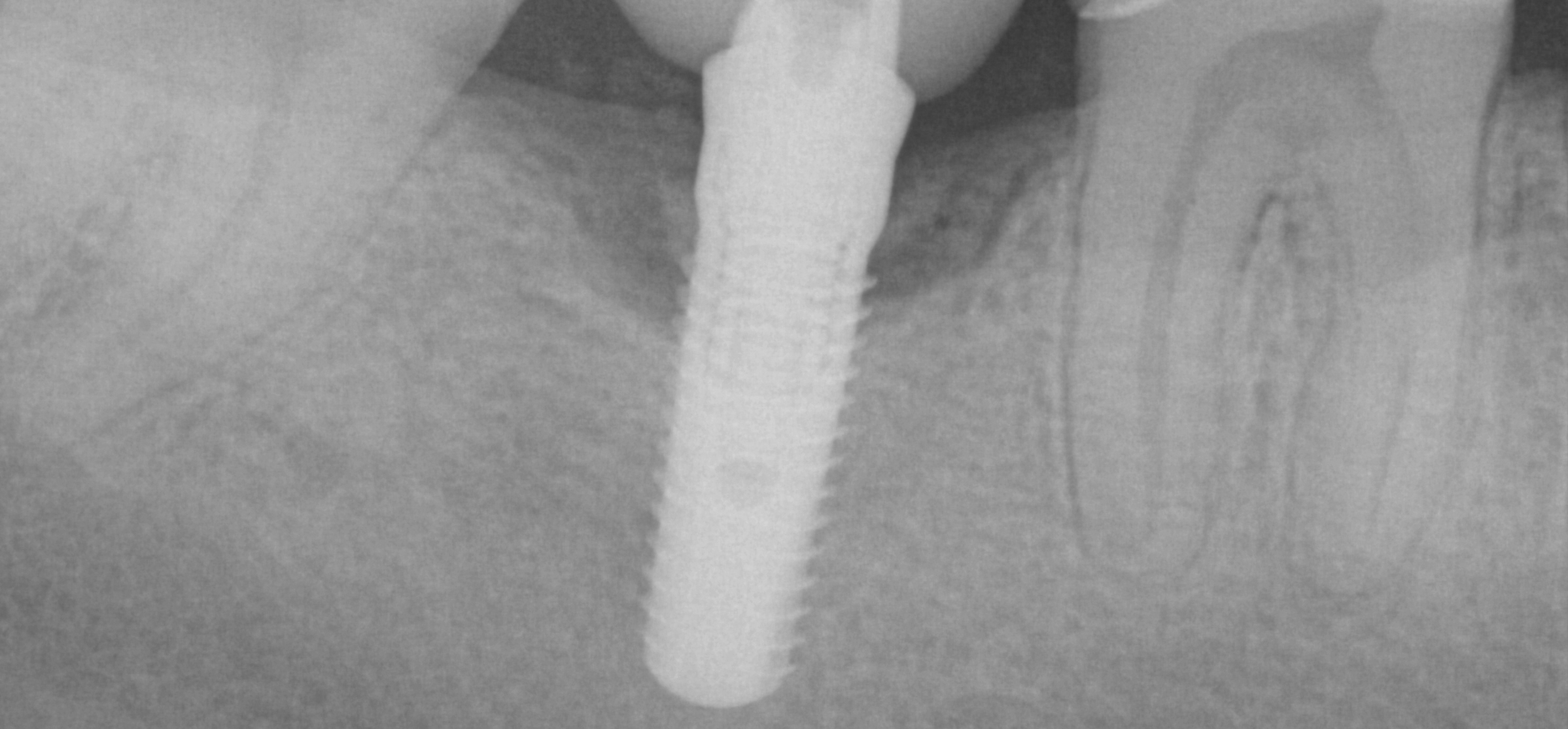
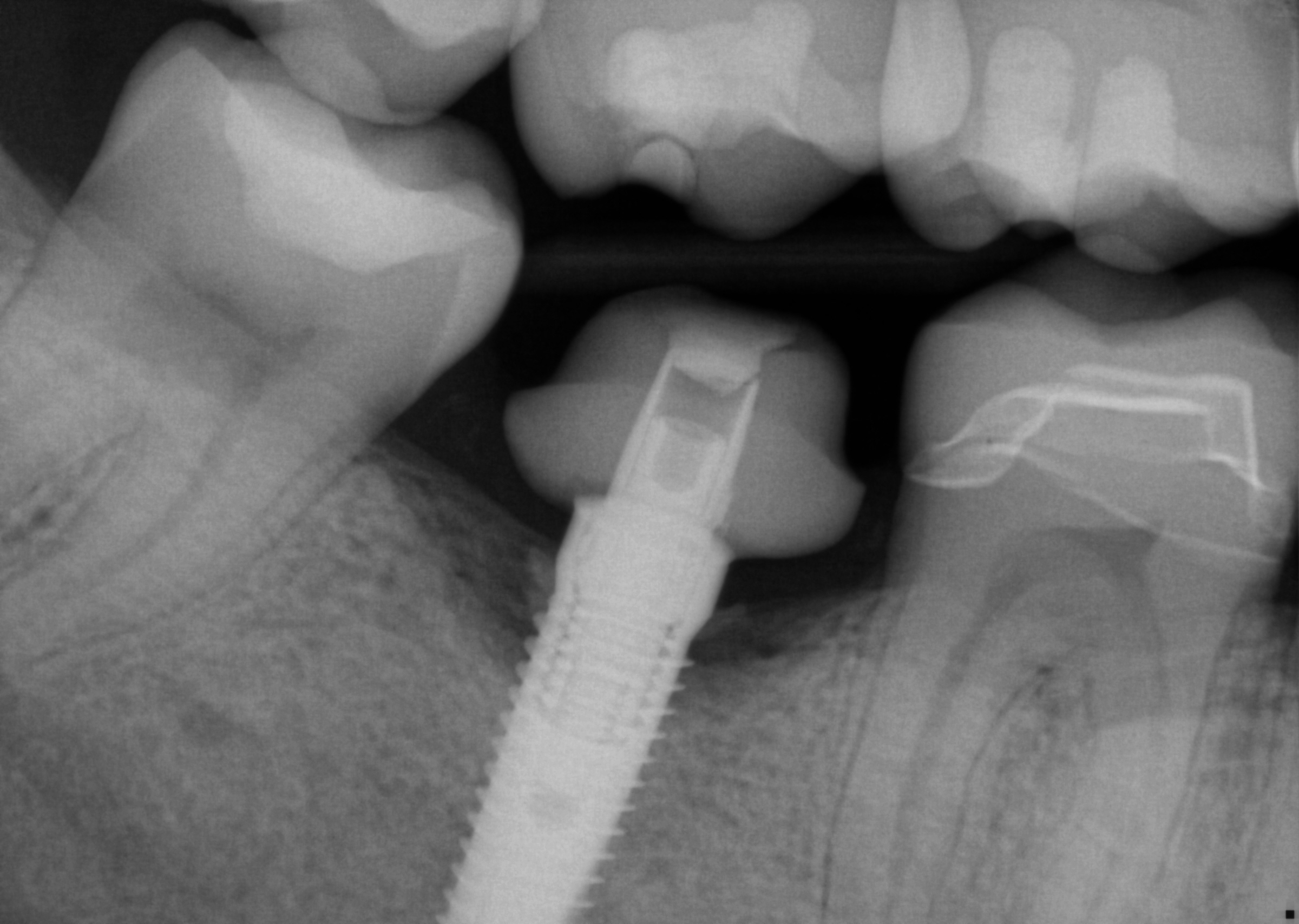
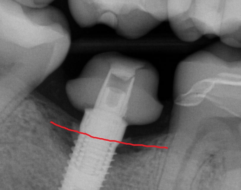
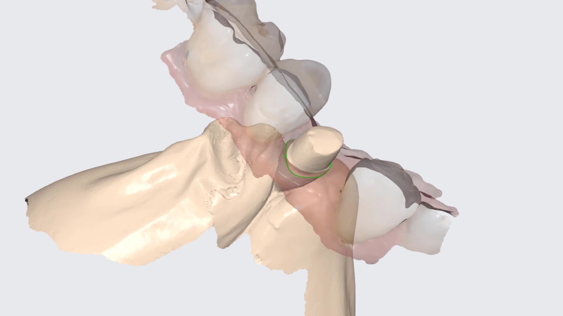
This implant was initially placed at the crest of the ridge in 2012. its placement did not allow proper emergence profile and subsequently lost bone around the head of the implant by 2015. The lack of blood flow to the crest of the bone at the cortical plate certainly could have contributed to its demise. By 2015, the bacteria trap forced a new restoration to be placed which also ultimately lead to chronic inflammation due to improper contours. The implant itself was well integrated and the decision was made to remove the top 3-5 mm of the implant and treat it as a cast post and core.
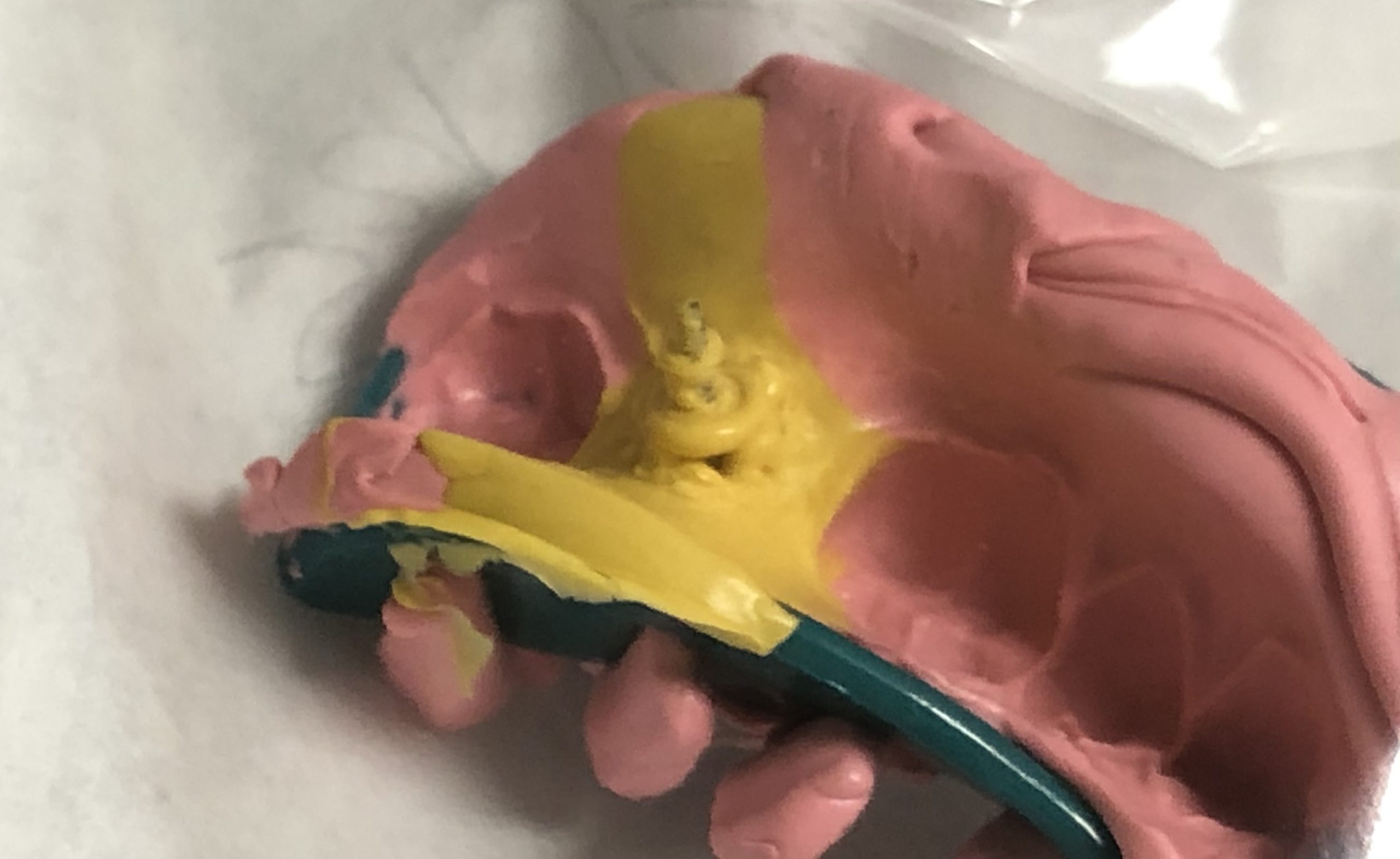
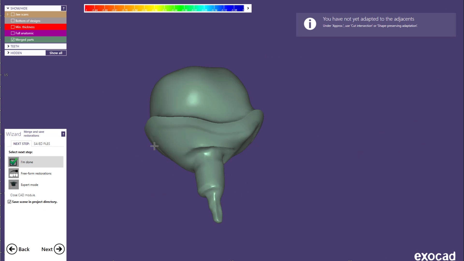
You can see how the Medit i700 was used to mix intra-oral scans with a PVS impression to capture the fixture. Advanced users can utilize the stl of the implant itself to fabricate a post and core digitally.
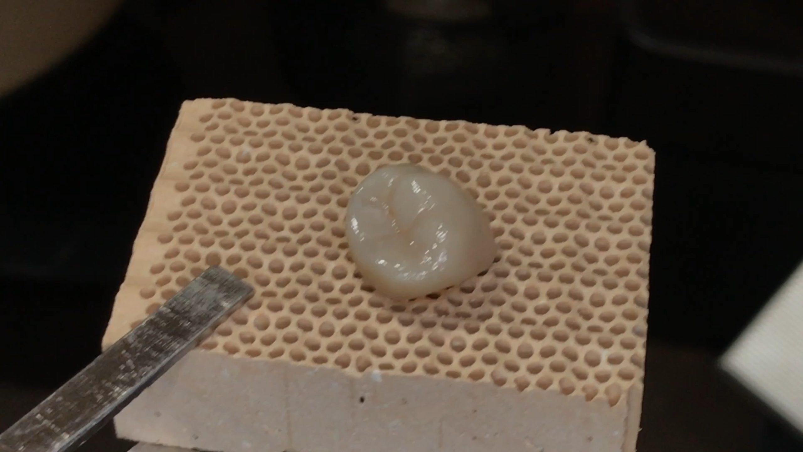
The post and core and the emax restoration were returned for delivery. The emax was not crystalized until after the abutment was cemented and then it was tried in by itself to assure proper contours and contacts. The great feature of lithium dissilicate is that you can add contact and glaze at the same time, which was required here as the mesial contact was weaker than desired after some minor adjustments


 the patient was sedated and intubated for the case so we could not keep track of the bite. Instead, we imaged all 30 prepared teeth and used medit compare / design to digitally mount them to the wax ups. In the link provided you can download the models and relate them to each other […]